 Movie
Movie Controller

[English] 日本語
 Yorodumi
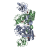
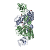
- PDB-6asl: Crystal Structure of Flavin monooxygenase CmoJ (earlier YtnJ) bou... -
+ Open data

- Basic information
| Entry | Database: PDB / ID: 6asl | ||||||
|---|---|---|---|---|---|---|---|
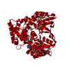
| Title | Crystal Structure of Flavin monooxygenase CmoJ (earlier YtnJ) bound with FMN | ||||||
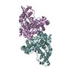
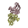
 Components Components | Putative monooxygenase MoxC | ||||||
 Keywords Keywords |  FLAVOPROTEIN / Flavin monooxygenase FLAVOPROTEIN / Flavin monooxygenase | ||||||
| Function / homology |  Function and homology informationOxidoreductases; Acting on paired donors, with incorporation or reduction of molecular oxygen; With reduced flavin or flavoprotein as one donor, and incorporation of one atom of oxygen into the other donor / oxidoreductase activity, acting on paired donors, with incorporation or reduction of molecular oxygen / monooxygenase activity Function and homology informationOxidoreductases; Acting on paired donors, with incorporation or reduction of molecular oxygen; With reduced flavin or flavoprotein as one donor, and incorporation of one atom of oxygen into the other donor / oxidoreductase activity, acting on paired donors, with incorporation or reduction of molecular oxygen / monooxygenase activitySimilarity search - Function | ||||||
| Biological species |  Bacillus subtilis (bacteria) Bacillus subtilis (bacteria) | ||||||
| Method | X-RAY DIFFRACTION / SYNCHROTRON / MOLECULAR REPLACEMENT / Resolution: 1.9 Å | ||||||
 Authors Authors | Bhandari, D.M. / Zhao, B. / Li, P. / Begley, T.P. | ||||||
| Funding support |  United States, 1items United States, 1items
| ||||||
 Citation Citation | Journal: To Be Published Title: Flavin mediated Pummerer type rearrangement in cysteine salvage pathway Authors: Bhandari, D.M. / Krishnamoorthy, K. / Zhao, B. / Li, P. / Begley, T.P. | ||||||
| History |
|
- Structure visualization
| Structure viewer | Molecule: MolmilJmol/JSmol |
|---|
- Downloads & links
-Download
| PDBx/mmCIF format | 6asl.cif.gz | 264.1 KB | Display | PDBx/mmCIF format |
|---|---|---|---|---|
| PDB format | pdb6asl.ent.gz | 215.5 KB | Display | PDB format |
| PDBx/mmJSON format | 6asl.json.gz | Tree view | PDBx/mmJSON format | |
| Others |  Other downloads Other downloads |
-Validation report
| Arichive directory | https://data.pdbj.org/pub/pdb/validation_reports/as/6aslftp://data.pdbj.org/pub/pdb/validation_reports/as/6asl | HTTPS FTP |
|---|
-Related structure data
| Related structure data |  6askC  1yw1S S: Starting model for refinement C: citing same article ( |
|---|---|
| Similar structure data |


-Links
 PDBj
PDBj
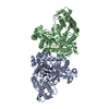
- Assembly
| Deposited unit | 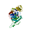
| ||||||||
|---|---|---|---|---|---|---|---|---|---|
| 1 | 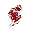
| ||||||||
| Unit cell |
|
-Components
| #1: Protein | Mass: 49669.395 Da / Num. of mol.: 1 Source method: isolated from a genetically manipulated source Source: (gene. exp.) Bacillus subtilis (strain 168) (bacteria)Strain: 168 / Gene: moxC, ytnJ, BSU29310 / Production host: Escherichia coli (E. coli) / Strain (production host): BL21(DE3) / References: UniProt: O34974 |
|---|---|
| #2: Chemical | ChemComp-FMN / Flavin mononucleotide  Mass: 456.344 Da / Num. of mol.: 1 / Source method: obtained synthetically / Formula: C17H21N4O9P Mass: 456.344 Da / Num. of mol.: 1 / Source method: obtained synthetically / Formula: C17H21N4O9P |
| #3: Chemical | ChemComp-LFN / Lumiflavin  Mass: 256.260 Da / Num. of mol.: 1 / Source method: obtained synthetically / Formula: C13H12N4O2 Mass: 256.260 Da / Num. of mol.: 1 / Source method: obtained synthetically / Formula: C13H12N4O2 |
| #4: Water | ChemComp-HOH / Water Mass: 18.015 Da / Num. of mol.: 216 / Source method: isolated from a natural source / Formula: H2O Mass: 18.015 Da / Num. of mol.: 216 / Source method: isolated from a natural source / Formula: H2O |
-Experimental details
-Experiment
| Experiment | Method: X-RAY DIFFRACTION / Number of used crystals: 1 |
|---|
- Sample preparation
| Crystal | Density Matthews: 3.72 Å3/Da / Density % sol: 66.94 % |
|---|---|
| Crystal grow | Temperature: 277 K / Method: vapor diffusion, hanging drop / pH: 7.5 Details: 50mM HEPES pH 7.5, 200 mM KCl, 29%-33% pentaerythritol propoxylate (5/4 PO/OH) |
-Data collection
| Diffraction | Mean temperature: 100 K |
|---|---|
| Diffraction source | Source: SYNCHROTRON / Site: APS / Beamline: 19-ID / Wavelength: 0.925 Å |
| Detector | Type: DECTRIS PILATUS 2M / Detector: PIXEL / Date: Jun 30, 2017 |
| Radiation | Protocol: SINGLE WAVELENGTH / Monochromatic (M) / Laue (L): M / Scattering type: x-ray |
| Radiation wavelength | Wavelength: 0.925 Å / Relative weight: 1 |
| Reflection | Resolution: 1.9→38.943 Å / Num. obs: 59542 / % possible obs: 100 % / Redundancy: 6.8 % / Rmerge(I) obs: 0.14 / Net I/σ(I): 16.14 |
| Reflection shell | Resolution: 1.9→1.97 Å / Redundancy: 5.4 % / Rmerge(I) obs: 1.029 / Mean I/σ(I) obs: 1 / % possible all: 99.83 |
- Processing
| Software |
| ||||||||||||||||||||||||||||||||||||||||||||||||||||||||||||||||||||||||||||||||||||||||||||||||||||||||||||||||||||||||||||||||||||||||||||||||||||||||||||||||||||||||||||||||||||||||||||||||||||||||
|---|---|---|---|---|---|---|---|---|---|---|---|---|---|---|---|---|---|---|---|---|---|---|---|---|---|---|---|---|---|---|---|---|---|---|---|---|---|---|---|---|---|---|---|---|---|---|---|---|---|---|---|---|---|---|---|---|---|---|---|---|---|---|---|---|---|---|---|---|---|---|---|---|---|---|---|---|---|---|---|---|---|---|---|---|---|---|---|---|---|---|---|---|---|---|---|---|---|---|---|---|---|---|---|---|---|---|---|---|---|---|---|---|---|---|---|---|---|---|---|---|---|---|---|---|---|---|---|---|---|---|---|---|---|---|---|---|---|---|---|---|---|---|---|---|---|---|---|---|---|---|---|---|---|---|---|---|---|---|---|---|---|---|---|---|---|---|---|---|---|---|---|---|---|---|---|---|---|---|---|---|---|---|---|---|---|---|---|---|---|---|---|---|---|---|---|---|---|---|---|---|---|
| Refinement | Method to determine structure: MOLECULAR REPLACEMENT Starting model: 1YW1 Resolution: 1.9→38.94 Å / SU ML: 0.18 / Cross valid method: FREE R-VALUE / σ(F): 1.34 / Phase error: 21.18
| ||||||||||||||||||||||||||||||||||||||||||||||||||||||||||||||||||||||||||||||||||||||||||||||||||||||||||||||||||||||||||||||||||||||||||||||||||||||||||||||||||||||||||||||||||||||||||||||||||||||||
| Solvent computation | Shrinkage radii: 0.9 Å / VDW probe radii: 1.11 Å | ||||||||||||||||||||||||||||||||||||||||||||||||||||||||||||||||||||||||||||||||||||||||||||||||||||||||||||||||||||||||||||||||||||||||||||||||||||||||||||||||||||||||||||||||||||||||||||||||||||||||
| Refinement step | Cycle: LAST / Resolution: 1.9→38.94 Å
| ||||||||||||||||||||||||||||||||||||||||||||||||||||||||||||||||||||||||||||||||||||||||||||||||||||||||||||||||||||||||||||||||||||||||||||||||||||||||||||||||||||||||||||||||||||||||||||||||||||||||
| Refine LS restraints |
| ||||||||||||||||||||||||||||||||||||||||||||||||||||||||||||||||||||||||||||||||||||||||||||||||||||||||||||||||||||||||||||||||||||||||||||||||||||||||||||||||||||||||||||||||||||||||||||||||||||||||
| LS refinement shell |
| ||||||||||||||||||||||||||||||||||||||||||||||||||||||||||||||||||||||||||||||||||||||||||||||||||||||||||||||||||||||||||||||||||||||||||||||||||||||||||||||||||||||||||||||||||||||||||||||||||||||||
| Refinement TLS params. | Method: refined / Refine-ID: X-RAY DIFFRACTION
| ||||||||||||||||||||||||||||||||||||||||||||||||||||||||||||||||||||||||||||||||||||||||||||||||||||||||||||||||||||||||||||||||||||||||||||||||||||||||||||||||||||||||||||||||||||||||||||||||||||||||
| Refinement TLS group |
|
