 Movie
Movie Controller

[English] 日本語
 Yorodumi
- PDB-6ar4: Crystal structure of PICK1 in complex with the small molecule inh... -
+ Open data

- Basic information
| Entry | Database: PDB / ID: 6ar4 | ||||||
|---|---|---|---|---|---|---|---|
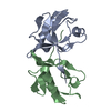
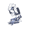
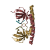
| Title | Crystal structure of PICK1 in complex with the small molecule inhibitor 1o | ||||||
 Components Components | PRKCA-binding protein | ||||||
 Keywords Keywords | Actin Binding protein / PDZ domain PDZ inhibitor | ||||||
| Function / homology |  Function and homology information Function and homology informationmembrane curvature sensor activity / postsynaptic early endosome / glial cell development / neuronal ion channel clustering / cellular response to decreased oxygen levels /  Arp2/3 complex binding / Trafficking of GluR2-containing AMPA receptors / epigenetic programming of gene expression / regulation of Arp2/3 complex-mediated actin nucleation / negative regulation of Arp2/3 complex-mediated actin nucleation ...membrane curvature sensor activity / postsynaptic early endosome / glial cell development / neuronal ion channel clustering / cellular response to decreased oxygen levels / Arp2/3 complex binding / Trafficking of GluR2-containing AMPA receptors / epigenetic programming of gene expression / regulation of Arp2/3 complex-mediated actin nucleation / negative regulation of Arp2/3 complex-mediated actin nucleation / monoamine transport / genomic imprinting / protein kinase C-activating G protein-coupled receptor signaling pathway / retrograde vesicle-mediated transport, Golgi to endoplasmic reticulum / dendritic spine organization / long-term synaptic depression / dendritic spine maintenance / receptor clustering / regulation of insulin secretion / positive regulation of receptor internalization / cellular response to glucose starvation / trans-Golgi network membrane / protein kinase C binding / G protein-coupled receptor binding / Cell surface interactions at the vascular wall / intracellular protein transport / phospholipid binding / endocytic vesicle membrane / actin filament binding / synaptic vesicle / presynaptic membrane / postsynaptic density / cytoskeleton / neuron projection / protein domain specific binding / protein phosphorylation / signaling receptor binding / synapse / perinuclear region of cytoplasm / Golgi apparatus / identical protein binding / metal ion binding / plasma membrane / cytosol / cytoplasm Arp2/3 complex binding / Trafficking of GluR2-containing AMPA receptors / epigenetic programming of gene expression / regulation of Arp2/3 complex-mediated actin nucleation / negative regulation of Arp2/3 complex-mediated actin nucleation ...membrane curvature sensor activity / postsynaptic early endosome / glial cell development / neuronal ion channel clustering / cellular response to decreased oxygen levels / Arp2/3 complex binding / Trafficking of GluR2-containing AMPA receptors / epigenetic programming of gene expression / regulation of Arp2/3 complex-mediated actin nucleation / negative regulation of Arp2/3 complex-mediated actin nucleation / monoamine transport / genomic imprinting / protein kinase C-activating G protein-coupled receptor signaling pathway / retrograde vesicle-mediated transport, Golgi to endoplasmic reticulum / dendritic spine organization / long-term synaptic depression / dendritic spine maintenance / receptor clustering / regulation of insulin secretion / positive regulation of receptor internalization / cellular response to glucose starvation / trans-Golgi network membrane / protein kinase C binding / G protein-coupled receptor binding / Cell surface interactions at the vascular wall / intracellular protein transport / phospholipid binding / endocytic vesicle membrane / actin filament binding / synaptic vesicle / presynaptic membrane / postsynaptic density / cytoskeleton / neuron projection / protein domain specific binding / protein phosphorylation / signaling receptor binding / synapse / perinuclear region of cytoplasm / Golgi apparatus / identical protein binding / metal ion binding / plasma membrane / cytosol / cytoplasmSimilarity search - Function | ||||||
| Biological species |  Homo sapiens (human) Homo sapiens (human) | ||||||
| Method | X-RAY DIFFRACTION / SYNCHROTRON / MOLECULAR REPLACEMENT / Resolution: 1.69 Å | ||||||
 Authors Authors | Marcotte, D. | ||||||
 Citation Citation | Journal: Sci Rep / Year: 2018 Title: Potent PDZ-Domain PICK1 Inhibitors that Modulate Amyloid Beta-Mediated Synaptic Dysfunction. Authors: Lin, E.Y.S. / Silvian, L.F. / Marcotte, D.J. / Banos, C.C. / Jow, F. / Chan, T.R. / Arduini, R.M. / Qian, F. / Baker, D.P. / Bergeron, C. / Hession, C.A. / Huganir, R.L. / Borenstein, C.F. / ...Authors: Lin, E.Y.S. / Silvian, L.F. / Marcotte, D.J. / Banos, C.C. / Jow, F. / Chan, T.R. / Arduini, R.M. / Qian, F. / Baker, D.P. / Bergeron, C. / Hession, C.A. / Huganir, R.L. / Borenstein, C.F. / Enyedy, I. / Zou, J. / Rohde, E. / Wittmann, M. / Kumaravel, G. / Rhodes, K.J. / Scannevin, R.H. / Dunah, A.W. / Guckian, K.M. | ||||||
| History |
|
- Structure visualization
| Structure viewer | Molecule: MolmilJmol/JSmol |
|---|
- Downloads & links
-Download
| PDBx/mmCIF format | 6ar4.cif.gz | 57.5 KB | Display | PDBx/mmCIF format |
|---|---|---|---|---|
| PDB format | pdb6ar4.ent.gz | 38.8 KB | Display | PDB format |
| PDBx/mmJSON format | 6ar4.json.gz | Tree view | PDBx/mmJSON format | |
| Others |  Other downloads Other downloads |
-Validation report
| Arichive directory | https://data.pdbj.org/pub/pdb/validation_reports/ar/6ar4ftp://data.pdbj.org/pub/pdb/validation_reports/ar/6ar4 | HTTPS FTP |
|---|
-Related structure data
| Related structure data | 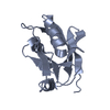 3hpkS S: Starting model for refinement |
|---|---|
| Similar structure data |


-Links
 PDBj
PDBj
- Assembly
| Deposited unit | 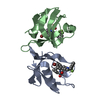
| ||||||||
|---|---|---|---|---|---|---|---|---|---|
| 1 |
| ||||||||
| Unit cell |
|
-Components
| #1: Protein | Mass: 13540.405 Da / Num. of mol.: 2 / Fragment: PDZ domain Source method: isolated from a genetically manipulated source Source: (gene. exp.) Homo sapiens (human) / Gene: PICK1, PRKCABP / Production host:  Escherichia coli (E. coli) / References: UniProt: Q9NRD5 Escherichia coli (E. coli) / References: UniProt: Q9NRD5#2: Chemical |   Mass: 553.411 Da / Num. of mol.: 2 / Source method: obtained synthetically / Formula: C26H28BrF3N2O3 Mass: 553.411 Da / Num. of mol.: 2 / Source method: obtained synthetically / Formula: C26H28BrF3N2O3#3: Water | ChemComp-HOH / | Water Mass: 18.015 Da / Num. of mol.: 247 / Source method: isolated from a natural source / Formula: H2O Mass: 18.015 Da / Num. of mol.: 247 / Source method: isolated from a natural source / Formula: H2O |
|---|
-Experimental details
-Experiment
| Experiment | Method: X-RAY DIFFRACTION / Number of used crystals: 1 |
|---|
- Sample preparation
| Crystal | Density Matthews: 2.44 Å3/Da / Density % sol: 49.61 % |
|---|---|
| Crystal grow | Temperature: 298 K / Method: vapor diffusion, sitting drop / pH: 6.5 / Details: 25% PEG3350, 0.1M BisTRIS pH 6.4 |
-Data collection
| Diffraction | Mean temperature: 100 K | ||||||||||||||||||||||||
|---|---|---|---|---|---|---|---|---|---|---|---|---|---|---|---|---|---|---|---|---|---|---|---|---|---|
| Diffraction source | Source: SYNCHROTRON / Site: APS  / Beamline: 31-ID / Wavelength: 0.98 Å / Beamline: 31-ID / Wavelength: 0.98 Å | ||||||||||||||||||||||||
| Detector | Type: MARMOSAIC 225 mm CCD / Detector: CCD / Date: Mar 2, 2012 | ||||||||||||||||||||||||
| Radiation | Protocol: SINGLE WAVELENGTH / Monochromatic (M) / Laue (L): M / Scattering type: x-ray | ||||||||||||||||||||||||
| Radiation wavelength | Wavelength: 0.98 Å / Relative weight: 1 | ||||||||||||||||||||||||
| Reflection | Resolution: 1.69→47.03 Å / Num. obs: 28410 / % possible obs: 99.1 % / Redundancy: 5.1 % / CC1/2: 0.999 / Rmerge(I) obs: 0.062 / Rpim(I) all: 0.03 / Rrim(I) all: 0.069 / Net I/σ(I): 16.4 | ||||||||||||||||||||||||
| Reflection shell | Diffraction-ID: 1
|
- Processing
| Software |
| ||||||||||||||||||||||||||||||||||||||||||||||||||||||||||||
|---|---|---|---|---|---|---|---|---|---|---|---|---|---|---|---|---|---|---|---|---|---|---|---|---|---|---|---|---|---|---|---|---|---|---|---|---|---|---|---|---|---|---|---|---|---|---|---|---|---|---|---|---|---|---|---|---|---|---|---|---|---|
| Refinement | Method to determine structure: MOLECULAR REPLACEMENT Starting model: 3HPK Resolution: 1.69→25.63 Å / Cor.coef. Fo:Fc: 0.969 / Cor.coef. Fo:Fc free: 0.952 / SU B: 0.362 / SU ML: 0.014 / Cross valid method: THROUGHOUT / σ(F): 0 / ESU R: 0.016 / ESU R Free: 0.017
| ||||||||||||||||||||||||||||||||||||||||||||||||||||||||||||
| Solvent computation | Ion probe radii: 0.8 Å / Shrinkage radii: 0.8 Å / VDW probe radii: 1.2 Å | ||||||||||||||||||||||||||||||||||||||||||||||||||||||||||||
| Displacement parameters | Biso max: 108.95 Å2 / Biso mean: 28.638 Å2 / Biso min: 15.81 Å2
| ||||||||||||||||||||||||||||||||||||||||||||||||||||||||||||
| Refinement step | Cycle: final / Resolution: 1.69→25.63 Å
| ||||||||||||||||||||||||||||||||||||||||||||||||||||||||||||
| Refine LS restraints |
| ||||||||||||||||||||||||||||||||||||||||||||||||||||||||||||
| LS refinement shell | Resolution: 1.69→1.734 Å / Rfactor Rfree error: 0 / Total num. of bins used: 20
|
