 Movie
Movie Controller


+ Open data

- Basic information
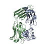
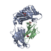
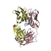
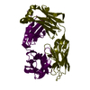
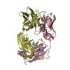
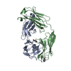
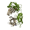
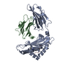
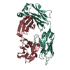
| Entry | Database: PDB / ID: 6apc | ||||||
|---|---|---|---|---|---|---|---|
| Title | Crystal Structure of Infant Antibody ADI-19425 | ||||||
 Components Components |
| ||||||
 Keywords Keywords |  IMMUNE SYSTEM / viral fusion glycoprotein / immunoglobulin / respiratory syncytial virus IMMUNE SYSTEM / viral fusion glycoprotein / immunoglobulin / respiratory syncytial virus | ||||||
| Function / homology |  Function and homology information Function and homology informationImmunoglobulin V-Type / Immunoglobulin V-set domain / Immunoglobulin V-set domain / Immunoglobulin/major histocompatibility complex, conserved site / Immunoglobulin subtype / Immunoglobulins and major histocompatibility complex proteins signature. / Immunoglobulin / Immunoglobulin C-Type / Immunoglobulin C1-set / Immunoglobulin C1-set domain ...Immunoglobulin V-Type / Immunoglobulin V-set domain / Immunoglobulin V-set domain / Immunoglobulin/major histocompatibility complex, conserved site / Immunoglobulin subtype / Immunoglobulins and major histocompatibility complex proteins signature. / Immunoglobulin / Immunoglobulin C-Type / Immunoglobulin C1-set / Immunoglobulin C1-set domain / Ig-like domain profile. / Immunoglobulin-like domain / Immunoglobulin-like domain superfamily / Immunoglobulins / Immunoglobulin-like fold / Immunoglobulin-like / Sandwich / Mainly BetaSimilarity search - Domain/homology | ||||||
| Biological species |  Homo sapiens (human) Homo sapiens (human) | ||||||
| Method | X-RAY DIFFRACTION / SYNCHROTRON / MOLECULAR REPLACEMENT / Resolution: 1.7 Å | ||||||
 Authors Authors | Gilman, M.S.A. / McLellan, J.S. | ||||||
 Citation Citation | Journal: Immunity / Year: 2018 Title: Infants Infected with Respiratory Syncytial Virus Generate Potent Neutralizing Antibodies that Lack Somatic Hypermutation. Authors: Goodwin, E. / Gilman, M.S.A. / Wrapp, D. / Chen, M. / Ngwuta, J.O. / Moin, S.M. / Bai, P. / Sivasubramanian, A. / Connor, R.I. / Wright, P.F. / Graham, B.S. / McLellan, J.S. / Walker, L.M. | ||||||
| History |
|
- Structure visualization
| Structure viewer | Molecule: MolmilJmol/JSmol |
|---|
- Downloads & links
-Download
| PDBx/mmCIF format | 6apc.cif.gz | 191.8 KB | Display | PDBx/mmCIF format |
|---|---|---|---|---|
| PDB format | pdb6apc.ent.gz | 148.3 KB | Display | PDB format |
| PDBx/mmJSON format | 6apc.json.gz | Tree view | PDBx/mmJSON format | |
| Others |  Other downloads Other downloads |
-Validation report
| Arichive directory | https://data.pdbj.org/pub/pdb/validation_reports/ap/6apcftp://data.pdbj.org/pub/pdb/validation_reports/ap/6apc | HTTPS FTP |
|---|
-Related structure data
| Related structure data |  6apbC  6apdC 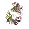 3eyqS 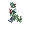 3h42S S: Starting model for refinement C: citing same article ( |
|---|---|
| Similar structure data |

-Links
 PDBj
PDBj

- Assembly
| Deposited unit | 
| ||||||||
|---|---|---|---|---|---|---|---|---|---|
| 1 |
| ||||||||
| Unit cell |
|
-Components
| #1: Antibody | Mass: 23881.695 Da / Num. of mol.: 1 Source method: isolated from a genetically manipulated source Source: (gene. exp.) Homo sapiens (human) / Gene: HEL-214 / Cell line (production host): FreeStyle 293-F / Production host: Homo sapiens (human) | ||
|---|---|---|---|
| #2: Antibody | Mass: 22955.275 Da / Num. of mol.: 1 Source method: isolated from a genetically manipulated source Source: (gene. exp.) Homo sapiens (human) / Gene: IGL@ / Cell (production host): FreeStyle 293-F / Production host: Homo sapiens (human) / References: UniProt: Q6GMX4 | ||
| #3: Chemical | Sulfate  Mass: 96.063 Da / Num. of mol.: 2 / Source method: obtained synthetically / Formula: SO4 Mass: 96.063 Da / Num. of mol.: 2 / Source method: obtained synthetically / Formula: SO4#4: Water | ChemComp-HOH / | Water Mass: 18.015 Da / Num. of mol.: 692 / Source method: isolated from a natural source / Formula: H2O Mass: 18.015 Da / Num. of mol.: 692 / Source method: isolated from a natural source / Formula: H2O |
-Experimental details
-Experiment
| Experiment | Method: X-RAY DIFFRACTION / Number of used crystals: 1 |
|---|
- Sample preparation
| Crystal | Density Matthews: 2.74 Å3/Da / Density % sol: 55.09 % |
|---|---|
| Crystal grow | Temperature: 293 K / Method: vapor diffusion, sitting drop / pH: 6.5 Details: 50 nL of ADI-19425 Fab at 8.78 mg/ml, 50 nL of crystal seed solution, and 100 nL of reservoir solution containing 1.5 M Ammonium sulfate, 0.1 M sodium chloride, and 0.1 M BisTris pH 6.5 |
-Data collection
| Diffraction | Mean temperature: 80 K | ||||||||||||||||||||||||
|---|---|---|---|---|---|---|---|---|---|---|---|---|---|---|---|---|---|---|---|---|---|---|---|---|---|
| Diffraction source | Source: SYNCHROTRON / Site: APS  / Beamline: 19-BM / Wavelength: 0.9792 Å / Beamline: 19-BM / Wavelength: 0.9792 Å | ||||||||||||||||||||||||
| Detector | Type: ADSC QUANTUM 210r / Detector: CCD / Date: Dec 18, 2015 | ||||||||||||||||||||||||
| Radiation | Protocol: SINGLE WAVELENGTH / Monochromatic (M) / Laue (L): M / Scattering type: x-ray | ||||||||||||||||||||||||
| Radiation wavelength | Wavelength: 0.9792 Å / Relative weight: 1 | ||||||||||||||||||||||||
| Reflection | Resolution: 1.7→26.07 Å / Num. obs: 57347 / % possible obs: 99.9 % / Redundancy: 6.8 % / Biso Wilson estimate: 14.07 Å2 / CC1/2: 0.998 / Rmerge(I) obs: 0.069 / Rpim(I) all: 0.028 / Rrim(I) all: 0.075 / Net I/σ(I): 16.8 | ||||||||||||||||||||||||
| Reflection shell | Diffraction-ID: 1
|
- Processing
| Software |
| ||||||||||||||||||||||||||||||||||||||||||||||||||||||||||||||||||||||||||||||||||||||||||||||||||||||||||||||||||||||||||||||||||||||||||||||||||||||||||
|---|---|---|---|---|---|---|---|---|---|---|---|---|---|---|---|---|---|---|---|---|---|---|---|---|---|---|---|---|---|---|---|---|---|---|---|---|---|---|---|---|---|---|---|---|---|---|---|---|---|---|---|---|---|---|---|---|---|---|---|---|---|---|---|---|---|---|---|---|---|---|---|---|---|---|---|---|---|---|---|---|---|---|---|---|---|---|---|---|---|---|---|---|---|---|---|---|---|---|---|---|---|---|---|---|---|---|---|---|---|---|---|---|---|---|---|---|---|---|---|---|---|---|---|---|---|---|---|---|---|---|---|---|---|---|---|---|---|---|---|---|---|---|---|---|---|---|---|---|---|---|---|---|---|---|---|
| Refinement | Method to determine structure: MOLECULAR REPLACEMENT Starting model: 3EYQ, 3H42 Resolution: 1.7→26.07 Å / SU ML: 0.17 / Cross valid method: FREE R-VALUE / σ(F): 1.33 / Phase error: 18.07
| ||||||||||||||||||||||||||||||||||||||||||||||||||||||||||||||||||||||||||||||||||||||||||||||||||||||||||||||||||||||||||||||||||||||||||||||||||||||||||
| Solvent computation | Shrinkage radii: 0.9 Å / VDW probe radii: 1.11 Å | ||||||||||||||||||||||||||||||||||||||||||||||||||||||||||||||||||||||||||||||||||||||||||||||||||||||||||||||||||||||||||||||||||||||||||||||||||||||||||
| Displacement parameters | Biso max: 53.18 Å2 / Biso mean: 18.44 Å2 / Biso min: 6.12 Å2 | ||||||||||||||||||||||||||||||||||||||||||||||||||||||||||||||||||||||||||||||||||||||||||||||||||||||||||||||||||||||||||||||||||||||||||||||||||||||||||
| Refinement step | Cycle: final / Resolution: 1.7→26.07 Å
| ||||||||||||||||||||||||||||||||||||||||||||||||||||||||||||||||||||||||||||||||||||||||||||||||||||||||||||||||||||||||||||||||||||||||||||||||||||||||||
| Refine LS restraints |
| ||||||||||||||||||||||||||||||||||||||||||||||||||||||||||||||||||||||||||||||||||||||||||||||||||||||||||||||||||||||||||||||||||||||||||||||||||||||||||
| LS refinement shell | Refine-ID: X-RAY DIFFRACTION / Rfactor Rfree error: 0 / Total num. of bins used: 21
| ||||||||||||||||||||||||||||||||||||||||||||||||||||||||||||||||||||||||||||||||||||||||||||||||||||||||||||||||||||||||||||||||||||||||||||||||||||||||||
| Refinement TLS params. | Method: refined / Refine-ID: X-RAY DIFFRACTION
| ||||||||||||||||||||||||||||||||||||||||||||||||||||||||||||||||||||||||||||||||||||||||||||||||||||||||||||||||||||||||||||||||||||||||||||||||||||||||||
| Refinement TLS group |
|
