 Movie
Movie Controller

[English] 日本語
 Yorodumi
- PDB-6an6: Crystal structure of Escherichia coli HPPK in complex with bisubs... -
+ Open data

- Basic information
| Entry | Database: PDB / ID: 6an6 | ||||||
|---|---|---|---|---|---|---|---|
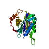
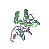
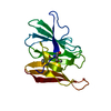
| Title | Crystal structure of Escherichia coli HPPK in complex with bisubstrate analogue inhibitor HP-72 | ||||||
 Components Components | 2-amino-4-hydroxy-6-hydroxymethyldihydropteridine pyrophosphokinase | ||||||
 Keywords Keywords | TRANSFERASE/INHIBITOR / alpha beta / TRANSFERASE-INHIBITOR complex | ||||||
| Function / homology |  Function and homology information Function and homology information2-amino-4-hydroxy-6-hydroxymethyldihydropteridine diphosphokinase / 2-amino-4-hydroxy-6-hydroxymethyldihydropteridine diphosphokinase activity / folic acid biosynthetic process / tetrahydrofolate biosynthetic process / kinase activity / magnesium ion binding / ATP binding Similarity search - Function | ||||||
| Biological species |  | ||||||
| Method |  X-RAY DIFFRACTION / SYNCHROTRON / MOLECULAR REPLACEMENT / Resolution: 2.3 Å X-RAY DIFFRACTION / SYNCHROTRON / MOLECULAR REPLACEMENT / Resolution: 2.3 Å | ||||||
 Authors Authors | Shaw, G.X. / Shi, G. / Ji, X. | ||||||
 Citation Citation | Journal: to be published Title: Bisubstrate analogue inhibitors of HPPK: Transition state mimetics Authors: Shaw, G.X. / Shi, G. / Ji, X. #1: Journal: J. Med. Chem. / Year: 2001Title: Bisubstrate analogue inhibitors of 6-hydroxymethyl-7,8-dihydropterin pyrophosphokinase: Synthesis and biochemical and crystallographic studies Authors: Shi, G. / Blaszczyk, J. / Ji, X. / Yan, H. #2: Journal: Bioorg. Med. Chem. / Year: 2012Title: Bisubstrate analogue inhibitors of 6-hydroxymethyl-7,8-dihydropterin pyrophosphokinase: New designnnnn with improved properties Authors: Shi, G. / Shaw, G. / Liang, Y.-H. / Subburamann, P. / Li, Y. / Wu, Y. / Yan, H. / Ji, X. | ||||||
| History |
|
- Structure visualization
| Structure viewer | Molecule: MolmilJmol/JSmol |
|---|
- Downloads & links
-Download
| PDBx/mmCIF format | 6an6.cif.gz | 135.6 KB | Display | PDBx/mmCIF format |
|---|---|---|---|---|
| PDB format | pdb6an6.ent.gz | 109.2 KB | Display | PDB format |
| PDBx/mmJSON format | 6an6.json.gz | Tree view | PDBx/mmJSON format | |
| Others |  Other downloads Other downloads |
-Validation report
| Summary document | 6an6_validation.pdf.gz | 997.8 KB | Display | wwPDB validaton report |
|---|---|---|---|---|
| Full document | 6an6_full_validation.pdf.gz | 1006.7 KB | Display | |
| Data in XML | 6an6_validation.xml.gz | 17 KB | Display | |
| Data in CIF | 6an6_validation.cif.gz | 22.7 KB | Display | |
| Arichive directory | https://data.pdbj.org/pub/pdb/validation_reports/an/6an6ftp://data.pdbj.org/pub/pdb/validation_reports/an/6an6 | HTTPS FTP |
-Related structure data
| Related structure data | 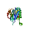 1eqmS S: Starting model for refinement |
|---|---|
| Similar structure data |


-Links
 PDBj
PDBj- Assembly
| Deposited unit | 
| ||||||||
|---|---|---|---|---|---|---|---|---|---|
| 1 | 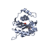
| ||||||||
| 2 | 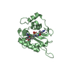
| ||||||||
| Unit cell |
|
-Components
| #1: Protein | Mass: 17966.535 Da / Num. of mol.: 2 Source method: isolated from a genetically manipulated source Source: (gene. exp.) Strain: K12 / Gene: folK, b0142, JW0138 / Plasmid: pDN2163 / Production host: Escherichia coli / Strain (production host): BL21-CodonPlus(DE3)-RIL References: UniProt: P26281, 2-amino-4-hydroxy-6-hydroxymethyldihydropteridine diphosphokinase #2: Chemical |   Mass: 682.649 Da / Num. of mol.: 2 / Source method: obtained synthetically / Formula: C24H35N12O8PS Mass: 682.649 Da / Num. of mol.: 2 / Source method: obtained synthetically / Formula: C24H35N12O8PS#3: Water | ChemComp-HOH / |  Mass: 18.015 Da / Num. of mol.: 142 / Source method: isolated from a natural source / Formula: H2O Mass: 18.015 Da / Num. of mol.: 142 / Source method: isolated from a natural source / Formula: H2O |
|---|
-Experimental details
-Experiment
| Experiment | Method: X-RAY DIFFRACTION / Number of used crystals: 1 |
|---|
- Sample preparation
| Crystal | Density Matthews: 2.04 Å3/Da / Density % sol: 39.57 % / Mosaicity: 2.474 ° |
|---|---|
| Crystal grow | Temperature: 292 K / Method: vapor diffusion, sitting drop / pH: 7 / Details: PEG 3350, NaCl etc. |
-Data collection
| Diffraction | Mean temperature: 100 K |
|---|---|
| Diffraction source | Source: SYNCHROTRON / Site: APS  / Beamline: 22-ID / Wavelength: 1 Å / Beamline: 22-ID / Wavelength: 1 Å |
| Detector | Type: RAYONIX MX300-HS / Detector: CCD / Date: Aug 18, 2013 / Details: mirrors |
| Radiation | Monochromator: Double crystal / Protocol: SINGLE WAVELENGTH / Monochromatic (M) / Laue (L): M / Scattering type: x-ray |
| Radiation wavelength | Wavelength: 1 Å / Relative weight: 1 |
| Reflection | Resolution: 2→30 Å / Num. obs: 16573 / % possible obs: 84.6 % / Redundancy: 3.9 % / Biso Wilson estimate: 17.1 Å2 / Rmerge(I) obs: 0.079 / Net I/σ(I): 10.9 |
| Reflection shell | Resolution: 2→2.07 Å / Redundancy: 2.6 % / Rmerge(I) obs: 0.181 / % possible all: 74.4 |
- Processing
| Software |
| ||||||||||||||||||||||||||||||||||||||||||
|---|---|---|---|---|---|---|---|---|---|---|---|---|---|---|---|---|---|---|---|---|---|---|---|---|---|---|---|---|---|---|---|---|---|---|---|---|---|---|---|---|---|---|---|
| Refinement | Method to determine structure: MOLECULAR REPLACEMENT Starting model: 1EQM Resolution: 2.3→29.68 Å / SU ML: 0.26 / Cross valid method: FREE R-VALUE / σ(F): 1.53 / Phase error: 31.81
| ||||||||||||||||||||||||||||||||||||||||||
| Solvent computation | Shrinkage radii: 0.9 Å / VDW probe radii: 1.11 Å | ||||||||||||||||||||||||||||||||||||||||||
| Displacement parameters | Biso mean: 22.88 Å2 | ||||||||||||||||||||||||||||||||||||||||||
| Refinement step | Cycle: LAST / Resolution: 2.3→29.68 Å
| ||||||||||||||||||||||||||||||||||||||||||
| Refine LS restraints |
| ||||||||||||||||||||||||||||||||||||||||||
| LS refinement shell |
|
