- PDB-4l1n: Crystal structure of a putative conserved lipoprotein (NT01CX_115... -
+
Open data
ID or keywords:
Loading...
-
Basic information
Entry
Database: PDB / ID: 4l1n
Title
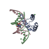
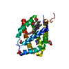
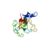
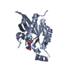
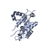
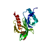
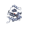
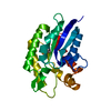
Crystal structure of a putative conserved lipoprotein (NT01CX_1156) from Clostridium novyi NT at 2.70 A resolution
Components
Conserved lipoprotein, putative
Keywords
STRUCTURAL GENOMICS / UNKNOWN FUNCTION / PF15525 family protein / DUF4652 / Joint Center for Structural Genomics / JCSG / Protein Structure Initiative / PSI-BIOLOGY
Function / homology
Uncharacterised protein PF15525, DUF4652 / Protein of unknown function DUF4652 / Domain of unknown function (DUF4652) / Lipocalin / membrane => GO:0016020 / Beta Barrel / Mainly Beta / Conserved lipoprotein, putative
Function and homology information
Biological species
Clostridium novyi (bacteria)
Method
X-RAY DIFFRACTION / SYNCHROTRON / SAD / Resolution: 2.7 Å
Mass: 18.015 Da / Num. of mol.: 10 / Source method: isolated from a natural source / Formula: H2O
Sequence details
THE CONSTRUCT (36-240) WAS EXPRESSED WITH A PURIFICATION TAG MGSDKIHHHHHHENLYFQG. THE TAG WAS ...THE CONSTRUCT (36-240) WAS EXPRESSED WITH A PURIFICATION TAG MGSDKIHHHHHHENLYFQG. THE TAG WAS REMOVED WITH TEV PROTEASE LEAVING ONLY A GLYCINE (0) FOLLOWED BY THE TARGET SEQUENCE.
-
Experimental details
-
Experiment
Experiment
Method: X-RAY DIFFRACTION / Number of used crystals: 1
-
Sample preparation
Crystal
Density Matthews: 5.54 Å3/Da / Density % sol: 77.79 %
Monochromator: double crystal Si(111) / Protocol: SINGLE WAVELENGTH / Monochromatic (M) / Laue (L): M / Scattering type: x-ray
Radiation wavelength
Wavelength: 0.9794 Å / Relative weight: 1
Reflection
Resolution: 2.7→44.915 Å / Num. obs: 14440 / % possible obs: 99.6 % / Observed criterion σ(I): -3 / Biso Wilson estimate: 81.523 Å2 / Rmerge(I) obs: 0.1 / Net I/σ(I): 11.41
Reflection shell
Rmerge(I) obs: 0.011 / Diffraction-ID: 1
Resolution (Å)
Highest resolution (Å)
Mean I/σ(I) obs
Num. measured obs
Num. unique obs
% possible all
2.7-2.77
1.8
5580
1073
99.7
2.77-2.85
2
5259
1013
99.4
2.85-2.93
2.5
5216
1008
99.9
2.93-3.02
3.6
4929
961
99.4
3.02-3.12
4.8
4795
946
99.1
3.12-3.23
5.7
4198
910
99.3
3.23-3.35
8.5
4392
891
99.3
3.35-3.49
10.6
4550
846
100
3.49-3.64
12.4
4413
829
100
3.64-3.82
14.9
4067
772
100
3.82-4.03
16.1
3860
742
99.9
4.03-4.27
18.2
3664
717
99.7
4.27-4.56
20.6
3211
658
98.9
4.56-4.93
20.1
2740
622
99.5
4.93-5.4
21.2
3028
574
99.8
5.4-6.04
20.4
2703
527
100
6.04-6.97
21.5
2345
466
99.6
6.97-8.54
23.6
1856
395
99.5
8.54-12.08
27.1
1434
310
98.4
12.08
28.4
857
180
97.3
-
Phasing
Phasing
Method: SAD
-
Processing
Software
Name
Version
Classification
NB
MolProbity
3beta29
modelbuilding
PDB_EXTRACT
3.1
dataextraction
SHELX
phasing
SHARP
phasing
XSCALE
March15, 2012
datascaling
BUSTER-TNT
2.10.0
refinement
XDS
datareduction
SHELXD
phasing
BUSTER
2.10.0
refinement
Refinement
Method to determine structure: SAD / Resolution: 2.7→44.915 Å / Cor.coef. Fo:Fc: 0.9128 / Cor.coef. Fo:Fc free: 0.9078 / Occupancy max: 1 / Occupancy min: 0.75 / Cross valid method: THROUGHOUT / σ(F): 0 Details: 1. A MET-INHIBITION PROTOCOL WAS USED FOR SELENOMETHIONINE INCORPORATION DURING PROTEIN EXPRESSION. THE OCCUPANCY OF THE SE ATOMS IN THE MSE RESIDUES WAS REDUCED TO 0.75 FOR THE REDUCED ...Details: 1. A MET-INHIBITION PROTOCOL WAS USED FOR SELENOMETHIONINE INCORPORATION DURING PROTEIN EXPRESSION. THE OCCUPANCY OF THE SE ATOMS IN THE MSE RESIDUES WAS REDUCED TO 0.75 FOR THE REDUCED SCATTERING POWER DUE TO PARTIAL S-MET INCORPORATION. 2. ATOM RECORD CONTAINS SUM OF TLS AND RESIDUAL B FACTORS. ANISOU RECORD CONTAINS SUM OF TLS AND RESIDUAL U FACTORS. 3. THE MAD PHASES WERE USED AS RESTRAINTS DURING REFINEMENT. 4. THE N-TERMINAL RESIDUES (36-79) ARE DISORDERED.
In the structure databanks used in Yorodumi, some data are registered as the other names, "COVID-19 virus" and "2019-nCoV". Here are the details of the virus and the list of structure data.
Jan 31, 2019. EMDB accession codes are about to change! (news from PDBe EMDB page)
EMDB accession codes are about to change! (news from PDBe EMDB page)
The allocation of 4 digits for EMDB accession codes will soon come to an end. Whilst these codes will remain in use, new EMDB accession codes will include an additional digit and will expand incrementally as the available range of codes is exhausted. The current 4-digit format prefixed with “EMD-” (i.e. EMD-XXXX) will advance to a 5-digit format (i.e. EMD-XXXXX), and so on. It is currently estimated that the 4-digit codes will be depleted around Spring 2019, at which point the 5-digit format will come into force.
The EM Navigator/Yorodumi systems omit the EMD- prefix.
Related info.:Q: What is EMD? / ID/Accession-code notation in Yorodumi/EM Navigator
Yorodumi is a browser for structure data from EMDB, PDB, SASBDB, etc.
This page is also the successor to EM Navigator detail page, and also detail information page/front-end page for Omokage search.
The word "yorodu" (or yorozu) is an old Japanese word meaning "ten thousand". "mi" (miru) is to see.
Related info.:EMDB / PDB / SASBDB / Comparison of 3 databanks / Yorodumi Search / Aug 31, 2016. New EM Navigator & Yorodumi / Yorodumi Papers / Jmol/JSmol / Function and homology information / Changes in new EM Navigator and Yorodumi
 Movie
Movie Controller

 Yorodumi
 Open data
 Basic information
 Components
 Keywords
 STRUCTURAL GENOMICS / UNKNOWN FUNCTION / PF15525 family protein / DUF4652 / Joint Center for Structural Genomics / JCSG /
  Function and homology information
 Authors
 Citation
 Structure visualization
 Downloads & links
 Other downloads
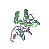

 PDBj
PDBj Assembly
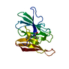
 Mass: 18.015 Da / Num. of mol.: 10 / Source method: isolated from a natural source / Formula: H2O
 Sample preparation
 / Beamline: BL12-2 / Wavelength: 0.9794
  Processing