 Movie
Movie Controller

[English] 日本語
 Yorodumi
- PDB-6ajc: Crystal structure of Trypanosoma cruzi cytosolic isocitrate dehyd... -
+ Open data

- Basic information
| Entry | Database: PDB / ID: 6ajc | ||||||
|---|---|---|---|---|---|---|---|
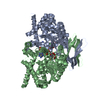
| Title | Crystal structure of Trypanosoma cruzi cytosolic isocitrate dehydrogenase in complex with NADP+, isocitrate and ca2+ | ||||||
 Components Components | Isocitrate dehydrogenase [NADP] | ||||||
 Keywords Keywords |  OXIDOREDUCTASE / Trypanosoma cruzi / metabolism / isocitrate dehydrogenase OXIDOREDUCTASE / Trypanosoma cruzi / metabolism / isocitrate dehydrogenase | ||||||
| Function / homology |  Function and homology information Function and homology informationisocitrate metabolic process / isocitrate dehydrogenase (NADP+) / isocitrate dehydrogenase (NADP+) activity / tricarboxylic acid cycle / NAD binding / magnesium ion bindingSimilarity search - Function | ||||||
| Biological species |  Trypanosoma cruzi (eukaryote) Trypanosoma cruzi (eukaryote) | ||||||
| Method | X-RAY DIFFRACTION / SYNCHROTRON / MOLECULAR REPLACEMENT / Resolution: 2.4 Å | ||||||
 Authors Authors | Wang, X. / Inaoka, D.K. / Shiba, T. / Balogun, E.O. / Ziebart, N. / Allman, S. / Watanabe, Y. / Nozaki, T. / Boshart, M. / Bringaud, F. ...Wang, X. / Inaoka, D.K. / Shiba, T. / Balogun, E.O. / Ziebart, N. / Allman, S. / Watanabe, Y. / Nozaki, T. / Boshart, M. / Bringaud, F. / Harada, S. / Kita, K. | ||||||
 Citation Citation | Journal: To Be Published Title: Biochemical characterization of a novel Trypanosoma brucei glycosomal isocitrate dehydrogenase with dual coenzyme specificity (NADP+/NAD+) Authors: Wang, X. / Inaoka, D.K. / Shiba, T. / Balogun, E.O. / Ziebart, N. / Allman, S. / Watanabe, Y. / Nozaki, T. / Boshart, M. / Bringaud, F. / Harada, S. / Kita, K. | ||||||
| History |
|
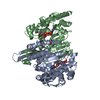
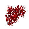
- Structure visualization
| Structure viewer | Molecule: MolmilJmol/JSmol |
|---|
- Downloads & links
-Download
| PDBx/mmCIF format | 6ajc.cif.gz | 646.2 KB | Display | PDBx/mmCIF format |
|---|---|---|---|---|
| PDB format | pdb6ajc.ent.gz | 538.5 KB | Display | PDB format |
| PDBx/mmJSON format | 6ajc.json.gz | Tree view | PDBx/mmJSON format | |
| Others |  Other downloads Other downloads |
-Validation report
| Arichive directory | https://data.pdbj.org/pub/pdb/validation_reports/aj/6ajcftp://data.pdbj.org/pub/pdb/validation_reports/aj/6ajc | HTTPS FTP |
|---|
-Related structure data
| Related structure data |  6aj6SC 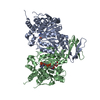 6aj8C 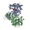 6ajaC 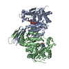 6ajbC S: Starting model for refinement C: citing same article ( |
|---|---|
| Similar structure data |


-Links
 PDBj
PDBj


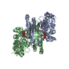
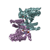
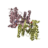
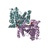
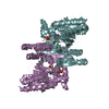
- Assembly
| Deposited unit | 
| ||||||||
|---|---|---|---|---|---|---|---|---|---|
| 1 | 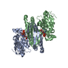
| ||||||||
| 2 | 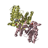
| ||||||||
| 3 | 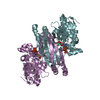
| ||||||||
| 4 | 
| ||||||||
| Unit cell |
|
-Components
| #1: Protein | Mass: 46881.633 Da / Num. of mol.: 8 / Mutation: M1S Source method: isolated from a genetically manipulated source Source: (gene. exp.) Trypanosoma cruzi (strain CL Brener) (eukaryote)Strain: CL Brener / Gene: Tc00.1047053506925.319 / Plasmid: pET-SUMO / Production host:  Escherichia coli (E. coli) Escherichia coli (E. coli)References: UniProt: Q4E4L7, isocitrate dehydrogenase (NADP+)#2: Chemical | ChemComp-NAP / Nicotinamide adenine dinucleotide phosphate  Mass: 743.405 Da / Num. of mol.: 8 / Source method: obtained synthetically / Formula: C21H28N7O17P3 Mass: 743.405 Da / Num. of mol.: 8 / Source method: obtained synthetically / Formula: C21H28N7O17P3#3: Chemical | ChemComp-CA /   Mass: 40.078 Da / Num. of mol.: 8 / Source method: obtained synthetically / Formula: Ca Mass: 40.078 Da / Num. of mol.: 8 / Source method: obtained synthetically / Formula: Ca#4: Chemical | ChemComp-ICT / Isocitric acid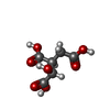  Mass: 192.124 Da / Num. of mol.: 8 / Source method: obtained synthetically / Formula: C6H8O7 Mass: 192.124 Da / Num. of mol.: 8 / Source method: obtained synthetically / Formula: C6H8O7#5: Water | ChemComp-HOH / | Water Mass: 18.015 Da / Num. of mol.: 121 / Source method: isolated from a natural source / Formula: H2O Mass: 18.015 Da / Num. of mol.: 121 / Source method: isolated from a natural source / Formula: H2O |
|---|
-Experimental details
-Experiment
| Experiment | Method: X-RAY DIFFRACTION / Number of used crystals: 1 |
|---|
- Sample preparation
| Crystal | Density Matthews: 2.49 Å3/Da / Density % sol: 50.6 % |
|---|---|
| Crystal grow | Temperature: 293 K / Method: vapor diffusion, sitting drop / pH: 6.5 / Details: 20% PEG 8000, 100mM imidazole/HCl pH6.5, 3% MPD |
-Data collection
| Diffraction | Mean temperature: 100 K |
|---|---|
| Diffraction source | Source: SYNCHROTRON / Site: SPring-8  / Beamline: BL44XU / Wavelength: 0.9 Å / Beamline: BL44XU / Wavelength: 0.9 Å |
| Detector | Type: RAYONIX MX300HE / Detector: CCD / Date: Apr 14, 2017 |
| Radiation | Protocol: SINGLE WAVELENGTH / Monochromatic (M) / Laue (L): M / Scattering type: x-ray |
| Radiation wavelength | Wavelength: 0.9 Å / Relative weight: 1 |
| Reflection | Resolution: 2.4→50 Å / Num. obs: 126184 / % possible obs: 86.7 % / Redundancy: 3.6 % / Rmerge(I) obs: 0.128 / Net I/σ(I): 8 |
| Reflection shell | Resolution: 2.4→2.44 Å / Rmerge(I) obs: 0.734 / Mean I/σ(I) obs: 1.02 / % possible all: 88.5 |
- Processing
| Software |
| ||||||||||||||||||||||||||||||||||||||||||||||||||||||||||||||||||||||||||||||||||||||||||||||||||||||||||||||||||||||||||||||||||||||||||||||||||||||||||||||||||||||||||||||||||||||
|---|---|---|---|---|---|---|---|---|---|---|---|---|---|---|---|---|---|---|---|---|---|---|---|---|---|---|---|---|---|---|---|---|---|---|---|---|---|---|---|---|---|---|---|---|---|---|---|---|---|---|---|---|---|---|---|---|---|---|---|---|---|---|---|---|---|---|---|---|---|---|---|---|---|---|---|---|---|---|---|---|---|---|---|---|---|---|---|---|---|---|---|---|---|---|---|---|---|---|---|---|---|---|---|---|---|---|---|---|---|---|---|---|---|---|---|---|---|---|---|---|---|---|---|---|---|---|---|---|---|---|---|---|---|---|---|---|---|---|---|---|---|---|---|---|---|---|---|---|---|---|---|---|---|---|---|---|---|---|---|---|---|---|---|---|---|---|---|---|---|---|---|---|---|---|---|---|---|---|---|---|---|---|---|
| Refinement | Method to determine structure: MOLECULAR REPLACEMENT Starting model: 6AJ6 Resolution: 2.4→20 Å / Cor.coef. Fo:Fc: 0.929 / Cor.coef. Fo:Fc free: 0.861 / SU B: 11.252 / SU ML: 0.263 / Cross valid method: THROUGHOUT / ESU R Free: 0.383 / Details: HYDROGENS HAVE BEEN ADDED IN THE RIDING POSITIONS
| ||||||||||||||||||||||||||||||||||||||||||||||||||||||||||||||||||||||||||||||||||||||||||||||||||||||||||||||||||||||||||||||||||||||||||||||||||||||||||||||||||||||||||||||||||||||
| Solvent computation | Ion probe radii: 0.8 Å / Shrinkage radii: 0.8 Å / VDW probe radii: 1.2 Å | ||||||||||||||||||||||||||||||||||||||||||||||||||||||||||||||||||||||||||||||||||||||||||||||||||||||||||||||||||||||||||||||||||||||||||||||||||||||||||||||||||||||||||||||||||||||
| Displacement parameters | Biso mean: 39.14 Å2
| ||||||||||||||||||||||||||||||||||||||||||||||||||||||||||||||||||||||||||||||||||||||||||||||||||||||||||||||||||||||||||||||||||||||||||||||||||||||||||||||||||||||||||||||||||||||
| Refinement step | Cycle: LAST / Resolution: 2.4→20 Å
| ||||||||||||||||||||||||||||||||||||||||||||||||||||||||||||||||||||||||||||||||||||||||||||||||||||||||||||||||||||||||||||||||||||||||||||||||||||||||||||||||||||||||||||||||||||||
| Refine LS restraints |
|