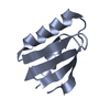
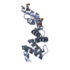
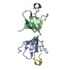
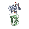
- PDB-6a71: Crystal Structure of Human ATP7B and TM Complex -
+
Open data
ID or keywords:
Loading...
-
Basic information
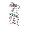
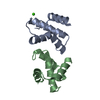
Entry
Database: PDB / ID: 6a71
Title
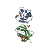
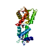
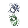
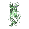
Crystal Structure of Human ATP7B and TM Complex
Components
ATP7B proteinWilson disease protein
Keywords
METAL TRANSPORT / Copper transporter protein
Function / homology
Function and homology information
regulation of biological quality / protein maturation by copper ion transfer / P-type divalent copper transporter activity / P-type monovalent copper transporter activity / P-type Cu+ transporter / copper ion export / copper ion import / copper ion transmembrane transporter activity / sequestering of calcium ion / copper ion transport ...regulation of biological quality / protein maturation by copper ion transfer / P-type divalent copper transporter activity / P-type monovalent copper transporter activity / P-type Cu+ transporter / copper ion export / copper ion import / copper ion transmembrane transporter activity / sequestering of calcium ion / copper ion transport / xenobiotic detoxification by transmembrane export across the plasma membrane / intracellular zinc ion homeostasis / response to copper ion / Ion transport by P-type ATPases / intracellular copper ion homeostasis / lactation / monoatomic ion transmembrane transport / trans-Golgi network membrane / establishment of localization in cell / trans-Golgi network / late endosome / copper ion binding / Golgi membrane / Golgi apparatus / ATP hydrolysis activity / mitochondrion / ATP binding / membrane / plasma membrane Similarity search - Function
Resolution: 1.6→43.87 Å / Cor.coef. Fo:Fc: 0.968 / Cor.coef. Fo:Fc free: 0.956 / SU B: 1.505 / SU ML: 0.053 / Cross valid method: THROUGHOUT / ESU R: 0.082 / ESU R Free: 0.083 / Stereochemistry target values: MAXIMUM LIKELIHOOD / Details: HYDROGENS HAVE BEEN ADDED IN THE RIDING POSITIONS
Rfactor
Num. reflection
% reflection
Selection details
Rfree
0.19537
1029
4.9 %
RANDOM
Rwork
0.16521
-
-
-
obs
0.16676
19928
97.76 %
-
Solvent computation
Ion probe radii: 0.8 Å / Shrinkage radii: 0.8 Å / VDW probe radii: 1.2 Å / Solvent model: MASK
 Movie
Movie Controller


 Open data
 Basic information
 Components
 Wilson disease protein
  Keywords
 Function and homology information
 Authors
 China, 1items
  Citation
 Structure visualization
 Downloads & links
 Other downloads

 PDBj
PDBj


 Assembly
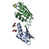


 Mass: 106.120 Da / Num. of mol.: 1 / Source method: obtained synthetically / Formula: C4H10O3
 Mass: 92.094 Da / Num. of mol.: 1 / Source method: obtained synthetically / Formula: C3H8O3
 Mass: 40.078 Da / Num. of mol.: 1 / Source method: obtained synthetically / Formula: Ca
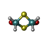
 Mass: 292.041 Da / Num. of mol.: 1 / Source method: obtained synthetically / Formula: H4Mo2O2S2
 Sample preparation
 Processing