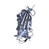
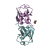
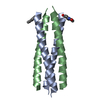
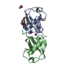
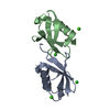
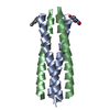
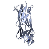
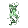
Journal: J Bacteriol / Year: 2013 Title: The structure of the CS1 pilus of enterotoxigenic Escherichia coli reveals structural polymorphism. Authors: Vitold E Galkin / Subramaniapillai Kolappan / Dixon Ng / ZuSheng Zong / Juliana Li / Xiong Yu / Edward H Egelman / Lisa Craig / Abstract: Enterotoxigenic Escherichia coli (ETEC) is a bacterial pathogen that causes diarrhea in children and travelers in developing countries. ETEC adheres to host epithelial cells in the small intestine ...Enterotoxigenic Escherichia coli (ETEC) is a bacterial pathogen that causes diarrhea in children and travelers in developing countries. ETEC adheres to host epithelial cells in the small intestine via a variety of different pili. The CS1 pilus is a prototype for a family of related pili, including the CFA/I pili, present on ETEC and other Gram-negative bacterial pathogens. These pili are assembled by an outer membrane usher protein that catalyzes subunit polymerization via donor strand complementation, in which the N terminus of each incoming pilin subunit fits into a hydrophobic groove in the terminal subunit, completing a β-sheet in the Ig fold. Here we determined a crystal structure of the CS1 major pilin subunit, CooA, to a 1.6-Å resolution. CooA is a globular protein with an Ig fold and is similar in structure to the CFA/I major pilin CfaB. We determined three distinct negative-stain electron microscopic reconstructions of the CS1 pilus and generated pseudoatomic-resolution pilus structures using the CooA crystal structure. CS1 pili adopt multiple structural states with differences in subunit orientations and packing. We propose that the structural perturbations are accommodated by flexibility in the N-terminal donor strand of CooA and by plasticity in interactions between exposed flexible loops on adjacent subunits. Our results suggest that CS1 and other pili of this class are extensible filaments that can be stretched in response to mechanical stress encountered during colonization.
Resolution: 1.6→19.67 Å / Cor.coef. Fo:Fc: 0.943 / Cor.coef. Fo:Fc free: 0.928 / SU B: 5.108 / SU ML: 0.088 / Cross valid method: THROUGHOUT / ESU R: 0.123 / ESU R Free: 0.115 / Stereochemistry target values: MAXIMUM LIKELIHOOD / Details: HYDROGENS HAVE BEEN ADDED IN THE RIDING POSITIONS
Rfactor
Num. reflection
% reflection
Selection details
Rfree
0.25057
1676
5 %
RANDOM
Rwork
0.22214
-
-
-
obs
0.22356
31826
94.94 %
-
all
-
35943
-
-
Solvent computation
Ion probe radii: 0.8 Å / Shrinkage radii: 0.8 Å / VDW probe radii: 1.2 Å / Solvent model: MASK
Displacement parameters
Biso mean: 22.257 Å2
Baniso -1
Baniso -2
Baniso -3
1-
0.24 Å2
-0.79 Å2
0.22 Å2
2-
-
1.73 Å2
-1.15 Å2
3-
-
-
-2.54 Å2
Refinement step
Cycle: LAST / Resolution: 1.6→19.67 Å
Protein
Nucleic acid
Ligand
Solvent
Total
Num. atoms
2212
0
6
156
2374
Refine LS restraints
Refine-ID
Type
Dev ideal
Dev ideal target
Number
X-RAY DIFFRACTION
r_bond_refined_d
0.008
0.019
2337
X-RAY DIFFRACTION
r_bond_other_d
0.001
0.02
2250
X-RAY DIFFRACTION
r_angle_refined_deg
1.374
1.962
3212
X-RAY DIFFRACTION
r_angle_other_deg
1.891
3
5207
X-RAY DIFFRACTION
r_dihedral_angle_1_deg
5.779
5
325
X-RAY DIFFRACTION
r_dihedral_angle_2_deg
35.686
27.403
77
X-RAY DIFFRACTION
r_dihedral_angle_3_deg
13.811
15
380
X-RAY DIFFRACTION
r_dihedral_angle_4_deg
15.23
15
2
X-RAY DIFFRACTION
r_chiral_restr
0.129
0.2
419
X-RAY DIFFRACTION
r_gen_planes_refined
0.005
0.021
2710
X-RAY DIFFRACTION
r_gen_planes_other
0.001
0.02
462
X-RAY DIFFRACTION
r_nbd_refined
X-RAY DIFFRACTION
r_nbd_other
X-RAY DIFFRACTION
r_nbtor_refined
0.163
0.2
1184
X-RAY DIFFRACTION
r_nbtor_other
X-RAY DIFFRACTION
r_xyhbond_nbd_refined
0.129
0.2
120
X-RAY DIFFRACTION
r_xyhbond_nbd_other
X-RAY DIFFRACTION
r_metal_ion_refined
0.077
0.2
1
X-RAY DIFFRACTION
r_metal_ion_other
X-RAY DIFFRACTION
r_symmetry_vdw_refined
0.292
0.2
16
X-RAY DIFFRACTION
r_symmetry_vdw_other
0.187
0.2
35
X-RAY DIFFRACTION
r_symmetry_hbond_refined
0.116
0.2
12
X-RAY DIFFRACTION
r_symmetry_hbond_other
X-RAY DIFFRACTION
r_symmetry_metal_ion_refined
X-RAY DIFFRACTION
r_symmetry_metal_ion_other
X-RAY DIFFRACTION
r_mcbond_it
0.563
1.5
1585
X-RAY DIFFRACTION
r_mcbond_other
0.127
1.5
637
X-RAY DIFFRACTION
r_mcangle_it
1.054
2
2612
X-RAY DIFFRACTION
r_scbond_it
1.741
3
756
X-RAY DIFFRACTION
r_scangle_it
2.864
4.5
600
X-RAY DIFFRACTION
r_rigid_bond_restr
X-RAY DIFFRACTION
r_sphericity_free
X-RAY DIFFRACTION
r_sphericity_bonded
LS refinement shell
Resolution: 1.6→1.641 Å / Total num. of bins used: 20
Rfactor
Num. reflection
% reflection
Rfree
0.294
118
-
Rwork
0.274
2225
-
obs
-
-
90.85 %
Refinement TLS params.
Method: refined / Refine-ID: X-RAY DIFFRACTION
ID
L11 (°2)
L12 (°2)
L13 (°2)
L22 (°2)
L23 (°2)
L33 (°2)
S11 (Å °)
S12 (Å °)
S13 (Å °)
S21 (Å °)
S22 (Å °)
S23 (Å °)
S31 (Å °)
S32 (Å °)
S33 (Å °)
T11 (Å2)
T12 (Å2)
T13 (Å2)
T22 (Å2)
T23 (Å2)
T33 (Å2)
Origin x (Å)
Origin y (Å)
Origin z (Å)
1
1.4186
-0.1691
0.1023
1.2359
-0.0863
2.1025
0.0325
0.0633
-0.0292
-0.028
-0.0243
0.0189
-0.2152
-0.0615
-0.0082
0.1182
0.0163
0.0272
0.0094
0.0288
0.2279
3.1448
2.4351
-15.1446
2
1.4421
0.2274
-0.0664
1.2852
0.2348
3.2138
0.0617
-0.0871
0.0161
0.0264
-0.0306
0.0096
0.1556
-0.0162
-0.0311
0.1153
0.0103
0.0134
0.0103
0.0251
0.2235
3.1955
-17.5715
-0.3097
Refinement TLS group
ID
Refine-ID
Refine TLS-ID
Auth asym-ID
Auth seq-ID
1
X-RAY DIFFRACTION
1
A
12 - 165
2
X-RAY DIFFRACTION
2
B
13 - 165
+
About Yorodumi
-
News
-
Feb 9, 2022. New format data for meta-information of EMDB entries
New format data for meta-information of EMDB entries
Version 3 of the EMDB header file is now the official format.
The previous official version 1.9 will be removed from the archive.
In the structure databanks used in Yorodumi, some data are registered as the other names, "COVID-19 virus" and "2019-nCoV". Here are the details of the virus and the list of structure data.
Jan 31, 2019. EMDB accession codes are about to change! (news from PDBe EMDB page)
EMDB accession codes are about to change! (news from PDBe EMDB page)
The allocation of 4 digits for EMDB accession codes will soon come to an end. Whilst these codes will remain in use, new EMDB accession codes will include an additional digit and will expand incrementally as the available range of codes is exhausted. The current 4-digit format prefixed with “EMD-” (i.e. EMD-XXXX) will advance to a 5-digit format (i.e. EMD-XXXXX), and so on. It is currently estimated that the 4-digit codes will be depleted around Spring 2019, at which point the 5-digit format will come into force.
The EM Navigator/Yorodumi systems omit the EMD- prefix.
Related info.:Q: What is EMD? / ID/Accession-code notation in Yorodumi/EM Navigator
Yorodumi is a browser for structure data from EMDB, PDB, SASBDB, etc.
This page is also the successor to EM Navigator detail page, and also detail information page/front-end page for Omokage search.
The word "yorodu" (or yorozu) is an old Japanese word meaning "ten thousand". "mi" (miru) is to see.
Related info.:EMDB / PDB / SASBDB / Comparison of 3 databanks / Yorodumi Search / Aug 31, 2016. New EM Navigator & Yorodumi / Yorodumi Papers / Jmol/JSmol / Function and homology information / Changes in new EM Navigator and Yorodumi
 Movie
Movie Controller

 Yorodumi
 Open data
 Basic information
 Components
 Keywords
 CELL ADHESION / CS1 pilus / COLONIZATION FACTOR /
  Function and homology information
 Authors
 Citation
 Structure visualization
 Downloads & links
 Other downloads


 PDBj
PDBj


 Assembly

 Mass: 69.085 Da / Num. of mol.: 1 / Source method: obtained synthetically / Formula: C3H5N2
 Mass: 22.990 Da / Num. of mol.: 1 / Source method: obtained synthetically / Formula: Na
 Mass: 18.015 Da / Num. of mol.: 156 / Source method: isolated from a natural source / Formula: H2O
 Sample preparation
 Processing