 Movie
Movie Controller

[English] 日本語
 Yorodumi
- PDB-6a6w: Crystal structure of fission yeast inner membrane protein Bqt4 in... -
+ Open data

- Basic information
| Entry | Database: PDB / ID: 6a6w | ||||||
|---|---|---|---|---|---|---|---|
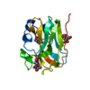
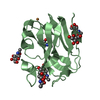
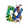
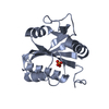
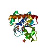
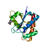
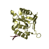
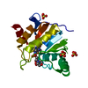
| Title | Crystal structure of fission yeast inner membrane protein Bqt4 in complex with Sad1 | ||||||
 Components Components |
| ||||||
 Keywords Keywords |  MEMBRANE PROTEIN / Chromosome organization / Inner nuclear membrane protein / Bouquet formation / Spindle pole boday MEMBRANE PROTEIN / Chromosome organization / Inner nuclear membrane protein / Bouquet formation / Spindle pole boday | ||||||
| Function / homology |  Function and homology information Function and homology informationold mitotic spindle pole body / new mitotic spindle pole body / spindle pole body-nuclear membrane anchor activity / mitotic spindle pole body localization / Sad1-Kms1 LINC complex / inner plaque of mitotic spindle pole body / centromere clustering at the mitotic interphase nuclear envelope / meiotic telomere tethering at nuclear periphery / telomere-nuclear envelope anchor activity / nuclear membrane complex Bqt3-Bqt4 ...old mitotic spindle pole body / new mitotic spindle pole body / spindle pole body-nuclear membrane anchor activity / mitotic spindle pole body localization / Sad1-Kms1 LINC complex / inner plaque of mitotic spindle pole body / centromere clustering at the mitotic interphase nuclear envelope / meiotic telomere tethering at nuclear periphery / telomere-nuclear envelope anchor activity / nuclear membrane complex Bqt3-Bqt4 / : / meiotic attachment of telomeric heterochromatin to spindle pole body / meiotic spindle pole body / mitotic telomere tethering at nuclear periphery / meiotic attachment of telomere to nuclear envelope / meiotic nuclear membrane microtubule tethering complex / mitotic spindle pole body / meiotic telomere clustering / nuclear envelope organization / interphase microtubule organizing center / nuclear inner membrane / protein-membrane adaptor activity / telomere organization / telomere maintenance / nuclear envelope / site of double-strand break / microtubule / cell division / DNA binding / nucleus / cytoplasmSimilarity search - Function | ||||||
| Biological species |  Schizosaccharomyces pombe (fission yeast) Schizosaccharomyces pombe (fission yeast) | ||||||
| Method | X-RAY DIFFRACTION / SYNCHROTRON / MOLECULAR REPLACEMENT / molecular replacement / Resolution: 2.601 Å | ||||||
 Authors Authors | Chen, Y. / Hu, C. | ||||||
 Citation Citation | Journal: Nucleic Acids Res. / Year: 2019 Title: Structural insights into chromosome attachment to the nuclear envelope by an inner nuclear membrane protein Bqt4 in fission yeast. Authors: Hu, C. / Inoue, H. / Sun, W. / Takeshita, Y. / Huang, Y. / Xu, Y. / Kanoh, J. / Chen, Y. | ||||||
| History |
|
- Structure visualization
| Structure viewer | Molecule: MolmilJmol/JSmol |
|---|
- Downloads & links
-Download
| PDBx/mmCIF format | 6a6w.cif.gz | 78 KB | Display | PDBx/mmCIF format |
|---|---|---|---|---|
| PDB format | pdb6a6w.ent.gz | 57.2 KB | Display | PDB format |
| PDBx/mmJSON format | 6a6w.json.gz | Tree view | PDBx/mmJSON format | |
| Others |  Other downloads Other downloads |
-Validation report
| Arichive directory | https://data.pdbj.org/pub/pdb/validation_reports/a6/6a6wftp://data.pdbj.org/pub/pdb/validation_reports/a6/6a6w | HTTPS FTP |
|---|
-Related structure data
| Related structure data |  5yc2C 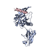 5ycaC 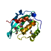 5ybxS S: Starting model for refinement C: citing same article ( |
|---|---|
| Similar structure data |


-Links
 PDBj
PDBj- Assembly
| Deposited unit | 
| ||||||||
|---|---|---|---|---|---|---|---|---|---|
| 1 |
| ||||||||
| Unit cell |
| ||||||||
| Components on special symmetry positions |
|
-Components
| #1: Protein | Mass: 15799.956 Da / Num. of mol.: 1 Source method: isolated from a genetically manipulated source Details: Bqt4 (residue 2 to 140) Source: (gene. exp.) Schizosaccharomyces pombe (strain 972 / ATCC 24843) (yeast)Strain: 972 / ATCC 24843 / Gene: bqt4, SPBC19C7.10 / Production host:  Escherichia coli BL21(DE3) (bacteria) / Strain (production host): BL21(DE3) / References: UniProt: O60158 Escherichia coli BL21(DE3) (bacteria) / Strain (production host): BL21(DE3) / References: UniProt: O60158 |
|---|---|
| #2: Protein/peptide | Mass: 1886.969 Da / Num. of mol.: 1 Source method: isolated from a genetically manipulated source Details: Sad1 residues 88 to 101 with three extra residues (GPL) from N-terminal tag Source: (gene. exp.) Schizosaccharomyces pombe (strain 972 / ATCC 24843) (yeast)Strain: 972 / ATCC 24843 / Gene: sad1, SPBC12D12.01, SPBC16H5.01c / Production host: Escherichia coli BL21(DE3) (bacteria) / Strain (production host): BL21(DE3) / References: UniProt: Q09825 |
| #3: Water | ChemComp-HOH / Water Mass: 18.015 Da / Num. of mol.: 15 / Source method: isolated from a natural source / Formula: H2O Mass: 18.015 Da / Num. of mol.: 15 / Source method: isolated from a natural source / Formula: H2O |
-Experimental details
-Experiment
| Experiment | Method: X-RAY DIFFRACTION / Number of used crystals: 1 |
|---|
- Sample preparation
| Crystal | Density Matthews: 6.42 Å3/Da / Density % sol: 80.84 % |
|---|---|
| Crystal grow | Temperature: 289 K / Method: vapor diffusion, sitting drop / pH: 7 / Details: 2.4M Sodium malonate, pH7.0 |
-Data collection
| Diffraction | Mean temperature: 100 K / Ambient temp details: liquid nitrogen | |||||||||||||||||||||||||||||||||||||||||||||||||||||||||||||||||||||||||||||||||||||||||||||||||||||||||||||||||||||||||||||||||||||||||||||||||||||||||||||||||||||||||||||||||||||||||||||
|---|---|---|---|---|---|---|---|---|---|---|---|---|---|---|---|---|---|---|---|---|---|---|---|---|---|---|---|---|---|---|---|---|---|---|---|---|---|---|---|---|---|---|---|---|---|---|---|---|---|---|---|---|---|---|---|---|---|---|---|---|---|---|---|---|---|---|---|---|---|---|---|---|---|---|---|---|---|---|---|---|---|---|---|---|---|---|---|---|---|---|---|---|---|---|---|---|---|---|---|---|---|---|---|---|---|---|---|---|---|---|---|---|---|---|---|---|---|---|---|---|---|---|---|---|---|---|---|---|---|---|---|---|---|---|---|---|---|---|---|---|---|---|---|---|---|---|---|---|---|---|---|---|---|---|---|---|---|---|---|---|---|---|---|---|---|---|---|---|---|---|---|---|---|---|---|---|---|---|---|---|---|---|---|---|---|---|---|---|---|---|
| Diffraction source | Source: SYNCHROTRON / Site: NFPSS  / Beamline: BL19U1 / Wavelength: 0.97775 Å / Beamline: BL19U1 / Wavelength: 0.97775 Å | |||||||||||||||||||||||||||||||||||||||||||||||||||||||||||||||||||||||||||||||||||||||||||||||||||||||||||||||||||||||||||||||||||||||||||||||||||||||||||||||||||||||||||||||||||||||||||||
| Detector | Type: DECTRIS PILATUS3 6M / Detector: PIXEL / Date: Jul 14, 2017 | |||||||||||||||||||||||||||||||||||||||||||||||||||||||||||||||||||||||||||||||||||||||||||||||||||||||||||||||||||||||||||||||||||||||||||||||||||||||||||||||||||||||||||||||||||||||||||||
| Radiation | Protocol: SINGLE WAVELENGTH / Monochromatic (M) / Laue (L): M / Scattering type: x-ray | |||||||||||||||||||||||||||||||||||||||||||||||||||||||||||||||||||||||||||||||||||||||||||||||||||||||||||||||||||||||||||||||||||||||||||||||||||||||||||||||||||||||||||||||||||||||||||||
| Radiation wavelength | Wavelength: 0.97775 Å / Relative weight: 1 | |||||||||||||||||||||||||||||||||||||||||||||||||||||||||||||||||||||||||||||||||||||||||||||||||||||||||||||||||||||||||||||||||||||||||||||||||||||||||||||||||||||||||||||||||||||||||||||
| Reflection | Resolution: 2.6→50 Å / Num. obs: 14717 / % possible obs: 100 % / Redundancy: 12.5 % / Rmerge(I) obs: 0.115 / Rpim(I) all: 0.033 / Rrim(I) all: 0.12 / Χ2: 1.412 / Net I/σ(I): 6.2 / Num. measured all: 184255 | |||||||||||||||||||||||||||||||||||||||||||||||||||||||||||||||||||||||||||||||||||||||||||||||||||||||||||||||||||||||||||||||||||||||||||||||||||||||||||||||||||||||||||||||||||||||||||||
| Reflection shell | Diffraction-ID: 1
|
-Phasing
| Phasing | Method: molecular replacement |
|---|
- Processing
| Software |
| ||||||||||||||||||||||||||||||||||||||||||||||||||||||||||||||||||||||||||||||||||||||||||||||||||||||||||||||||||||||||||||||||||||||||||||||||||||||||||||||||||||||||||||||||||||||||||||||||||||||||||||||||||||||||||||||||||||||||||||||||||||||||||
|---|---|---|---|---|---|---|---|---|---|---|---|---|---|---|---|---|---|---|---|---|---|---|---|---|---|---|---|---|---|---|---|---|---|---|---|---|---|---|---|---|---|---|---|---|---|---|---|---|---|---|---|---|---|---|---|---|---|---|---|---|---|---|---|---|---|---|---|---|---|---|---|---|---|---|---|---|---|---|---|---|---|---|---|---|---|---|---|---|---|---|---|---|---|---|---|---|---|---|---|---|---|---|---|---|---|---|---|---|---|---|---|---|---|---|---|---|---|---|---|---|---|---|---|---|---|---|---|---|---|---|---|---|---|---|---|---|---|---|---|---|---|---|---|---|---|---|---|---|---|---|---|---|---|---|---|---|---|---|---|---|---|---|---|---|---|---|---|---|---|---|---|---|---|---|---|---|---|---|---|---|---|---|---|---|---|---|---|---|---|---|---|---|---|---|---|---|---|---|---|---|---|---|---|---|---|---|---|---|---|---|---|---|---|---|---|---|---|---|---|---|---|---|---|---|---|---|---|---|---|---|---|---|---|---|---|---|---|---|---|---|---|---|---|---|---|---|---|---|---|---|---|
| Refinement | Method to determine structure: MOLECULAR REPLACEMENT Starting model: 5YBX Resolution: 2.601→43.846 Å / SU ML: 0.31 / Cross valid method: THROUGHOUT / σ(F): 1.38 / Phase error: 22.35 / Stereochemistry target values: ML
| ||||||||||||||||||||||||||||||||||||||||||||||||||||||||||||||||||||||||||||||||||||||||||||||||||||||||||||||||||||||||||||||||||||||||||||||||||||||||||||||||||||||||||||||||||||||||||||||||||||||||||||||||||||||||||||||||||||||||||||||||||||||||||
| Solvent computation | Shrinkage radii: 0.9 Å / VDW probe radii: 1.11 Å / Solvent model: FLAT BULK SOLVENT MODEL | ||||||||||||||||||||||||||||||||||||||||||||||||||||||||||||||||||||||||||||||||||||||||||||||||||||||||||||||||||||||||||||||||||||||||||||||||||||||||||||||||||||||||||||||||||||||||||||||||||||||||||||||||||||||||||||||||||||||||||||||||||||||||||
| Displacement parameters | Biso max: 147.43 Å2 / Biso mean: 66.396 Å2 / Biso min: 34.53 Å2 | ||||||||||||||||||||||||||||||||||||||||||||||||||||||||||||||||||||||||||||||||||||||||||||||||||||||||||||||||||||||||||||||||||||||||||||||||||||||||||||||||||||||||||||||||||||||||||||||||||||||||||||||||||||||||||||||||||||||||||||||||||||||||||
| Refinement step | Cycle: final / Resolution: 2.601→43.846 Å
| ||||||||||||||||||||||||||||||||||||||||||||||||||||||||||||||||||||||||||||||||||||||||||||||||||||||||||||||||||||||||||||||||||||||||||||||||||||||||||||||||||||||||||||||||||||||||||||||||||||||||||||||||||||||||||||||||||||||||||||||||||||||||||
| Refine LS restraints |
| ||||||||||||||||||||||||||||||||||||||||||||||||||||||||||||||||||||||||||||||||||||||||||||||||||||||||||||||||||||||||||||||||||||||||||||||||||||||||||||||||||||||||||||||||||||||||||||||||||||||||||||||||||||||||||||||||||||||||||||||||||||||||||
| LS refinement shell | Refine-ID: X-RAY DIFFRACTION / Rfactor Rfree error: 0 / Total num. of bins used: 6
| ||||||||||||||||||||||||||||||||||||||||||||||||||||||||||||||||||||||||||||||||||||||||||||||||||||||||||||||||||||||||||||||||||||||||||||||||||||||||||||||||||||||||||||||||||||||||||||||||||||||||||||||||||||||||||||||||||||||||||||||||||||||||||
| Refinement TLS params. | Method: refined / Refine-ID: X-RAY DIFFRACTION
| ||||||||||||||||||||||||||||||||||||||||||||||||||||||||||||||||||||||||||||||||||||||||||||||||||||||||||||||||||||||||||||||||||||||||||||||||||||||||||||||||||||||||||||||||||||||||||||||||||||||||||||||||||||||||||||||||||||||||||||||||||||||||||
| Refinement TLS group |
|
