 Movie
Movie Controller

[English] 日本語
 Yorodumi
- PDB-5zk3: Crystal structure of rationally thermostabilized M2 muscarinic ac... -
+ Open data

- Basic information
| Entry | Database: PDB / ID: 5zk3 | |||||||||||||||||||||
|---|---|---|---|---|---|---|---|---|---|---|---|---|---|---|---|---|---|---|---|---|---|---|
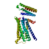
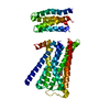
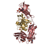
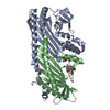
| Title | Crystal structure of rationally thermostabilized M2 muscarinic acetylcholine receptor bound with QNB | |||||||||||||||||||||
 Components Components | Muscarinic acetylcholine receptor M2,Apo-cytochrome b562,Muscarinic acetylcholine receptor M2 | |||||||||||||||||||||
 Keywords Keywords |  MEMBRANE PROTEIN/INHIBITOR / GPCR crystallography / rationally thermostabilized mutant / MEMBRANE PROTEIN-INHIBITOR complex MEMBRANE PROTEIN/INHIBITOR / GPCR crystallography / rationally thermostabilized mutant / MEMBRANE PROTEIN-INHIBITOR complex | |||||||||||||||||||||
| Function / homology |  Function and homology informationMuscarinic acetylcholine receptors / symmetric synapse / phospholipase C-activating G protein-coupled acetylcholine receptor signaling pathway / G protein-coupled acetylcholine receptor activity / regulation of smooth muscle contraction / cholinergic synapse / adenylate cyclase-inhibiting G protein-coupled acetylcholine receptor signaling pathway / G protein-coupled serotonin receptor activity / arrestin family protein binding / regulation of heart contraction ...Muscarinic acetylcholine receptors / symmetric synapse / phospholipase C-activating G protein-coupled acetylcholine receptor signaling pathway / G protein-coupled acetylcholine receptor activity / regulation of smooth muscle contraction / cholinergic synapse / adenylate cyclase-inhibiting G protein-coupled acetylcholine receptor signaling pathway / G protein-coupled serotonin receptor activity / arrestin family protein binding / regulation of heart contraction / G protein-coupled receptor signaling pathway, coupled to cyclic nucleotide second messenger / asymmetric synapse / axon terminus / presynaptic modulation of chemical synaptic transmission / clathrin-coated endocytic vesicle membrane / response to virus / adenylate cyclase-modulating G protein-coupled receptor signaling pathway / G protein-coupled acetylcholine receptor signaling pathway / Cargo recognition for clathrin-mediated endocytosis / presynaptic membrane / Clathrin-mediated endocytosis / nervous system development / G alpha (i) signalling events / chemical synaptic transmission / postsynaptic membrane / G protein-coupled receptor signaling pathway / neuronal cell body / dendrite / synapse / glutamatergic synapse / membrane / plasma membrane Function and homology informationMuscarinic acetylcholine receptors / symmetric synapse / phospholipase C-activating G protein-coupled acetylcholine receptor signaling pathway / G protein-coupled acetylcholine receptor activity / regulation of smooth muscle contraction / cholinergic synapse / adenylate cyclase-inhibiting G protein-coupled acetylcholine receptor signaling pathway / G protein-coupled serotonin receptor activity / arrestin family protein binding / regulation of heart contraction ...Muscarinic acetylcholine receptors / symmetric synapse / phospholipase C-activating G protein-coupled acetylcholine receptor signaling pathway / G protein-coupled acetylcholine receptor activity / regulation of smooth muscle contraction / cholinergic synapse / adenylate cyclase-inhibiting G protein-coupled acetylcholine receptor signaling pathway / G protein-coupled serotonin receptor activity / arrestin family protein binding / regulation of heart contraction / G protein-coupled receptor signaling pathway, coupled to cyclic nucleotide second messenger / asymmetric synapse / axon terminus / presynaptic modulation of chemical synaptic transmission / clathrin-coated endocytic vesicle membrane / response to virus / adenylate cyclase-modulating G protein-coupled receptor signaling pathway / G protein-coupled acetylcholine receptor signaling pathway / Cargo recognition for clathrin-mediated endocytosis / presynaptic membrane / Clathrin-mediated endocytosis / nervous system development / G alpha (i) signalling events / chemical synaptic transmission / postsynaptic membrane / G protein-coupled receptor signaling pathway / neuronal cell body / dendrite / synapse / glutamatergic synapse / membrane / plasma membraneSimilarity search - Function | |||||||||||||||||||||
| Biological species |  Homo sapiens (human) Homo sapiens (human) | |||||||||||||||||||||
| Method | X-RAY DIFFRACTION / SYNCHROTRON / MOLECULAR REPLACEMENT / Resolution: 2.6 Å | |||||||||||||||||||||
 Authors Authors | Suno, R. / Maeda, S. / Yasuda, S. / Yamashita, K. / Hirata, K. / Horita, S. / Tawaramoto, M.S. / Tsujimoto, H. / Murata, T. / Kinoshita, M. ...Suno, R. / Maeda, S. / Yasuda, S. / Yamashita, K. / Hirata, K. / Horita, S. / Tawaramoto, M.S. / Tsujimoto, H. / Murata, T. / Kinoshita, M. / Yamamoto, M. / Kobilka, B.K. / Iwata, S. / Kobayashi, T. | |||||||||||||||||||||
| Funding support |  Japan, Japan,  United States, 6items United States, 6items
| |||||||||||||||||||||
 Citation Citation | Journal: Nat. Chem. Biol. / Year: 2018 Title: Structural insights into the subtype-selective antagonist binding to the M2muscarinic receptor Authors: Suno, R. / Lee, S. / Maeda, S. / Yasuda, S. / Yamashita, K. / Hirata, K. / Horita, S. / Tawaramoto, M.S. / Tsujimoto, H. / Murata, T. / Kinoshita, M. / Yamamoto, M. / Kobilka, B.K. / ...Authors: Suno, R. / Lee, S. / Maeda, S. / Yasuda, S. / Yamashita, K. / Hirata, K. / Horita, S. / Tawaramoto, M.S. / Tsujimoto, H. / Murata, T. / Kinoshita, M. / Yamamoto, M. / Kobilka, B.K. / Vaidehi, N. / Iwata, S. / Kobayashi, T. | |||||||||||||||||||||
| History |
|
- Structure visualization
| Structure viewer | Molecule: MolmilJmol/JSmol |
|---|
- Downloads & links
-Download
| PDBx/mmCIF format | 5zk3.cif.gz | 164.7 KB | Display | PDBx/mmCIF format |
|---|---|---|---|---|
| PDB format | pdb5zk3.ent.gz | 135.8 KB | Display | PDB format |
| PDBx/mmJSON format | 5zk3.json.gz | Tree view | PDBx/mmJSON format | |
| Others |  Other downloads Other downloads |
-Validation report
| Arichive directory | https://data.pdbj.org/pub/pdb/validation_reports/zk/5zk3ftp://data.pdbj.org/pub/pdb/validation_reports/zk/5zk3 | HTTPS FTP |
|---|
-Related structure data
| Related structure data |  5yc8C  5zk8C 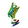 5zkbC  5zkcC  5xba 5xbb C: citing same article ( |
|---|---|
| Similar structure data | |
| Experimental dataset #1 | Data reference: 10.5281/zenodo.1094808 / Data set type: diffraction image data |
-Links
 PDBj
PDBj


- Assembly
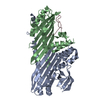
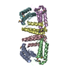
| Deposited unit | 
| ||||||||
|---|---|---|---|---|---|---|---|---|---|
| 1 |
| ||||||||
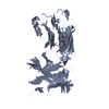
| Unit cell |
|
-Components
| #1: Protein | Mass: 47293.207 Da / Num. of mol.: 1 / Fragment: UNP residues 10-217,UNP residues 377-466 / Mutation: S110R Source method: isolated from a genetically manipulated source Source: (gene. exp.) Homo sapiens (human) / Gene: CHRM2 / Production host:   Spodoptera frugiperda (fall armyworm) / References: UniProt: P08172 Spodoptera frugiperda (fall armyworm) / References: UniProt: P08172 |
|---|---|
| #2: Chemical | ChemComp-QNB / (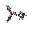  Mass: 337.412 Da / Num. of mol.: 1 / Source method: obtained synthetically / Formula: C21H23NO3 Mass: 337.412 Da / Num. of mol.: 1 / Source method: obtained synthetically / Formula: C21H23NO3 |
| #3: Water | ChemComp-HOH / Water Mass: 18.015 Da / Num. of mol.: 5 / Source method: isolated from a natural source / Formula: H2O Mass: 18.015 Da / Num. of mol.: 5 / Source method: isolated from a natural source / Formula: H2O |
-Experimental details
-Experiment
| Experiment | Method: X-RAY DIFFRACTION / Number of used crystals: 1 |
|---|
- Sample preparation
| Crystal | Density Matthews: 2.55 Å3/Da / Density % sol: 51.75 % |
|---|---|
| Crystal grow | Temperature: 293 K / Method: lipidic cubic phase Details: 50mM MES-NaOH pH 6.2-7.0, 26-32% PEG300, 300~500mM Ammonium Fluoride, 1% 1,2,3-heptanetriol, 0.5mM QNB and 5% DMSO |
-Data collection
| Diffraction | Mean temperature: 100 K |
|---|---|
| Diffraction source | Source: SYNCHROTRON / Site: SPring-8 / Beamline: BL32XU / Wavelength: 1 Å |
| Detector | Type: RAYONIX MX225-HS / Detector: CCD / Date: Jul 19, 2016 |
| Radiation | Protocol: SINGLE WAVELENGTH / Monochromatic (M) / Laue (L): M / Scattering type: x-ray |
| Radiation wavelength | Wavelength: 1 Å / Relative weight: 1 |
| Reflection | Resolution: 2.6→50 Å / Num. obs: 14817 / % possible obs: 99.9 % / Redundancy: 12.7 % / Net I/σ(I): 8.28 |
| Reflection shell | Resolution: 2.6→2.76 Å |
- Processing
| Software |
| ||||||||||||||||||||||||||||||||||||||||||||||||||||||||||||||||||||||||||||||||||||||||||||||||||||||||||||||||||||||||||||||||||||||||||||||||||||||
|---|---|---|---|---|---|---|---|---|---|---|---|---|---|---|---|---|---|---|---|---|---|---|---|---|---|---|---|---|---|---|---|---|---|---|---|---|---|---|---|---|---|---|---|---|---|---|---|---|---|---|---|---|---|---|---|---|---|---|---|---|---|---|---|---|---|---|---|---|---|---|---|---|---|---|---|---|---|---|---|---|---|---|---|---|---|---|---|---|---|---|---|---|---|---|---|---|---|---|---|---|---|---|---|---|---|---|---|---|---|---|---|---|---|---|---|---|---|---|---|---|---|---|---|---|---|---|---|---|---|---|---|---|---|---|---|---|---|---|---|---|---|---|---|---|---|---|---|---|---|---|---|
| Refinement | Method to determine structure: MOLECULAR REPLACEMENT / Resolution: 2.6→45.908 Å / SU ML: 0.36 / Cross valid method: FREE R-VALUE / σ(F): 1.37 / Phase error: 31.82
| ||||||||||||||||||||||||||||||||||||||||||||||||||||||||||||||||||||||||||||||||||||||||||||||||||||||||||||||||||||||||||||||||||||||||||||||||||||||
| Solvent computation | Shrinkage radii: 0.9 Å / VDW probe radii: 1.11 Å | ||||||||||||||||||||||||||||||||||||||||||||||||||||||||||||||||||||||||||||||||||||||||||||||||||||||||||||||||||||||||||||||||||||||||||||||||||||||
| Refinement step | Cycle: LAST / Resolution: 2.6→45.908 Å
| ||||||||||||||||||||||||||||||||||||||||||||||||||||||||||||||||||||||||||||||||||||||||||||||||||||||||||||||||||||||||||||||||||||||||||||||||||||||
| Refine LS restraints |
| ||||||||||||||||||||||||||||||||||||||||||||||||||||||||||||||||||||||||||||||||||||||||||||||||||||||||||||||||||||||||||||||||||||||||||||||||||||||
| LS refinement shell |
| ||||||||||||||||||||||||||||||||||||||||||||||||||||||||||||||||||||||||||||||||||||||||||||||||||||||||||||||||||||||||||||||||||||||||||||||||||||||
| Refinement TLS params. | Method: refined / Refine-ID: X-RAY DIFFRACTION
| ||||||||||||||||||||||||||||||||||||||||||||||||||||||||||||||||||||||||||||||||||||||||||||||||||||||||||||||||||||||||||||||||||||||||||||||||||||||
| Refinement TLS group |
|
