 Movie
Movie Controller


+ Open data

- Basic information
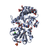
| Entry | Database: PDB / ID: 5zjq | ||||||
|---|---|---|---|---|---|---|---|
| Title | Structure of AbdB/Exd complex bound to a 'Red14' DNA sequence | ||||||
 Components Components |
| ||||||
 Keywords Keywords | TRANSCRIPTION/DNA / AbdB / Exd /  DNA / shape / specificity / TRANSCRIPTION / TRANSCRIPTION-DNA complex DNA / shape / specificity / TRANSCRIPTION / TRANSCRIPTION-DNA complex | ||||||
| Function / homology |  Function and homology information Function and homology informationgenital disc sexually dimorphic development / male pigmentation / negative regulation of cardioblast cell fate specification / sperm storage / negative regulation of salivary gland boundary specification / specification of segmental identity, abdomen / imaginal disc-derived female genitalia development / determination of genital disc primordium / external genitalia morphogenesis / salivary gland boundary specification ...genital disc sexually dimorphic development / male pigmentation / negative regulation of cardioblast cell fate specification / sperm storage / negative regulation of salivary gland boundary specification / specification of segmental identity, abdomen / imaginal disc-derived female genitalia development / determination of genital disc primordium / external genitalia morphogenesis / salivary gland boundary specification / gonadal mesoderm development / oenocyte development / : / female genitalia development / open tracheal system development / somatic muscle development / genital disc anterior/posterior pattern formation / spiracle morphogenesis, open tracheal system / polytene chromosome band / negative regulation of female receptivity / negative regulation of striated muscle tissue development / neuroendocrine cell differentiation / positive regulation of apoptotic process involved in morphogenesis / segment specification / eye development / regulation of cell fate specification / germ cell migration / male genitalia development / peripheral nervous system development / midgut development / transcription factor binding / neuron development / embryonic organ development / cis-regulatory region sequence-specific DNA binding / protein-DNA complex / transcription coregulator activity / animal organ morphogenesis / brain development / RNA polymerase II transcription regulator complex / male gonad development / DNA-binding transcription factor binding / transcription regulator complex / sequence-specific DNA binding / transcription coactivator activity / transcription cis-regulatory region binding / DNA-binding transcription factor activity, RNA polymerase II-specific / RNA polymerase II cis-regulatory region sequence-specific DNA binding / DNA-binding transcription factor activity / protein heterodimerization activity / regulation of transcription by RNA polymerase II / positive regulation of DNA-templated transcription / positive regulation of transcription by RNA polymerase II / DNA binding / nucleus / cytoplasmSimilarity search - Function | ||||||
| Biological species |  Drosophila melanogaster (fruit fly) Drosophila melanogaster (fruit fly) | ||||||
| Method | X-RAY DIFFRACTION / SYNCHROTRON / MOLECULAR REPLACEMENT / Resolution: 2.443 Å | ||||||
 Authors Authors | Baburajendran, N. / Zeiske, T. / Kaczynska, A. / Mann, R. / Honig, B. / Shapiro, L. / Palmer, A.G. | ||||||
 Citation Citation | Journal: Cell Rep / Year: 2018 Title: Intrinsic DNA Shape Accounts for Affinity Differences between Hox-Cofactor Binding Sites. Authors: Zeiske, T. / Baburajendran, N. / Kaczynska, A. / Brasch, J. / Palmer, A.G. / Shapiro, L. / Honig, B. / Mann, R.S. | ||||||
| History |
|
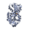
- Structure visualization
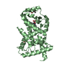
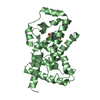
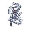
| Structure viewer | Molecule: MolmilJmol/JSmol |
|---|
- Downloads & links
-Download
| PDBx/mmCIF format | 5zjq.cif.gz | 105.6 KB | Display | PDBx/mmCIF format |
|---|---|---|---|---|
| PDB format | pdb5zjq.ent.gz | 76.6 KB | Display | PDB format |
| PDBx/mmJSON format | 5zjq.json.gz | Tree view | PDBx/mmJSON format | |
| Others |  Other downloads Other downloads |
-Validation report
| Arichive directory | https://data.pdbj.org/pub/pdb/validation_reports/zj/5zjqftp://data.pdbj.org/pub/pdb/validation_reports/zj/5zjq | HTTPS FTP |
|---|
-Related structure data
| Related structure data |  5zjrC 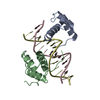 5zjsC 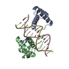 5zjtC  1b8iS S: Starting model for refinement C: citing same article ( |
|---|---|
| Similar structure data |


-Links
 PDBj
PDBj


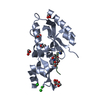
- Assembly
| Deposited unit | 
| ||||||||||
|---|---|---|---|---|---|---|---|---|---|---|---|
| 1 |
| ||||||||||
| Unit cell |
|
-Components
| #1: Protein | / Infraabdominal 7 / IAB-7 / P3 / PH189 Mass: 10195.751 Da / Num. of mol.: 1 Source method: isolated from a genetically manipulated source Source: (gene. exp.) Drosophila melanogaster (fruit fly) / Gene: Abd-B, CG10291 / Production host:  Escherichia coli (E. coli) / References: UniProt: P09087 Escherichia coli (E. coli) / References: UniProt: P09087 |
|---|---|
| #2: Protein | / Dpbx Mass: 8773.827 Da / Num. of mol.: 1 Source method: isolated from a genetically manipulated source Source: (gene. exp.) Drosophila melanogaster (fruit fly) / Gene: exd, CG8933 / Production host: Escherichia coli (E. coli) / References: UniProt: P40427 |
| #3: DNA chain | Mass: 4263.804 Da / Num. of mol.: 1 / Source method: obtained synthetically / Source: (synth.) Drosophila melanogaster (fruit fly) |
| #4: DNA chain | Mass: 4294.814 Da / Num. of mol.: 1 / Source method: obtained synthetically / Source: (synth.) Drosophila melanogaster (fruit fly) |
| #5: Water | ChemComp-HOH / Water Mass: 18.015 Da / Num. of mol.: 43 / Source method: isolated from a natural source / Formula: H2O Mass: 18.015 Da / Num. of mol.: 43 / Source method: isolated from a natural source / Formula: H2O |
-Experimental details
-Experiment
| Experiment | Method: X-RAY DIFFRACTION / Number of used crystals: 1 |
|---|
- Sample preparation
| Crystal | Density Matthews: 3.11 Å3/Da / Density % sol: 60.42 % |
|---|---|
| Crystal grow | Temperature: 293 K / Method: vapor diffusion Details: 200mM MgCl2, 0.1M Tris pH 5.8, 17.5% PEG 4000, 5% glycerol |
-Data collection
| Diffraction | Mean temperature: 100 K |
|---|---|
| Diffraction source | Source: SYNCHROTRON / Site: APS  / Beamline: 24-ID-E / Wavelength: 0.97918 Å / Beamline: 24-ID-E / Wavelength: 0.97918 Å |
| Detector | Type: ADSC QUANTUM 315 / Detector: CCD / Date: Nov 27, 2013 |
| Radiation | Protocol: SINGLE WAVELENGTH / Monochromatic (M) / Laue (L): M / Scattering type: x-ray |
| Radiation wavelength | Wavelength: 0.97918 Å / Relative weight: 1 |
| Reflection | Resolution: 2.443→29.942 Å / Num. obs: 12413 / % possible obs: 97.33 % / Redundancy: 3.4 % / Rmerge(I) obs: 0.065 / Net I/σ(I): 18.49 |
| Reflection shell | Resolution: 2.443→2.53 Å / Rmerge(I) obs: 0.539 / Num. unique obs: 980 |
- Processing
| Software |
| ||||||||||||||||||||||||||||||||||||||||||||||||||||||||||||||||||||||
|---|---|---|---|---|---|---|---|---|---|---|---|---|---|---|---|---|---|---|---|---|---|---|---|---|---|---|---|---|---|---|---|---|---|---|---|---|---|---|---|---|---|---|---|---|---|---|---|---|---|---|---|---|---|---|---|---|---|---|---|---|---|---|---|---|---|---|---|---|---|---|---|
| Refinement | Method to determine structure: MOLECULAR REPLACEMENT Starting model: 1B8I Resolution: 2.443→29.942 Å / SU ML: 0.33 / Cross valid method: THROUGHOUT / σ(F): 1.34 / Phase error: 29.34
| ||||||||||||||||||||||||||||||||||||||||||||||||||||||||||||||||||||||
| Solvent computation | Shrinkage radii: 0.9 Å / VDW probe radii: 1.11 Å | ||||||||||||||||||||||||||||||||||||||||||||||||||||||||||||||||||||||
| Displacement parameters | Biso max: 126.31 Å2 / Biso mean: 45.5612 Å2 / Biso min: 18.63 Å2 | ||||||||||||||||||||||||||||||||||||||||||||||||||||||||||||||||||||||
| Refinement step | Cycle: final / Resolution: 2.443→29.942 Å
| ||||||||||||||||||||||||||||||||||||||||||||||||||||||||||||||||||||||
| Refine LS restraints |
| ||||||||||||||||||||||||||||||||||||||||||||||||||||||||||||||||||||||
| LS refinement shell | Refine-ID: X-RAY DIFFRACTION / Rfactor Rfree error: 0 / Total num. of bins used: 9
| ||||||||||||||||||||||||||||||||||||||||||||||||||||||||||||||||||||||
| Refinement TLS params. | Method: refined / Origin x: -18.1278 Å / Origin y: 1.8931 Å / Origin z: 119.8547 Å
| ||||||||||||||||||||||||||||||||||||||||||||||||||||||||||||||||||||||
| Refinement TLS group | Selection details: all |
