 Movie
Movie Controller


+ Open data

- Basic information
| Entry | Database: PDB / ID: 1b8i | ||||||
|---|---|---|---|---|---|---|---|
| Title | STRUCTURE OF THE HOMEOTIC UBX/EXD/DNA TERNARY COMPLEX | ||||||
 Components Components |
| ||||||
 Keywords Keywords | TRANSCRIPTION/DNA /  DNA BINDING / HOMEODOMAIN / HOMEOTIC PROTEINS / DEVELOPMENT / SPECIFICITY / TRANSCRIPTION-DNA COMPLEX DNA BINDING / HOMEODOMAIN / HOMEOTIC PROTEINS / DEVELOPMENT / SPECIFICITY / TRANSCRIPTION-DNA COMPLEX | ||||||
| Function / homology |  Function and homology information Function and homology informationdorsal vessel aortic cell fate commitment / positive regulation of muscle organ development / anterior Malpighian tubule development / specification of animal organ identity / regulation of imaginal disc growth / salivary gland boundary specification / haltere development / oenocyte development / mesodermal cell fate specification / specification of segmental identity, thorax ...dorsal vessel aortic cell fate commitment / positive regulation of muscle organ development / anterior Malpighian tubule development / specification of animal organ identity / regulation of imaginal disc growth / salivary gland boundary specification / haltere development / oenocyte development / mesodermal cell fate specification / specification of segmental identity, thorax / open tracheal system development / imaginal disc-derived leg morphogenesis / somatic muscle development / muscle cell fate specification / polytene chromosome band / endoderm formation / eye development / regulation of cell fate specification / peripheral nervous system development / cell fate determination / anterior/posterior pattern specification / midgut development / transcription factor binding / neuron development / embryonic organ development / cis-regulatory region sequence-specific DNA binding / transcription repressor complex / transcription coregulator binding / protein-DNA complex / transcription coregulator activity / animal organ morphogenesis / brain development / RNA polymerase II transcription regulator complex / heart development / DNA-binding transcription factor binding / transcription regulator complex / sequence-specific DNA binding / transcription coactivator activity / DNA-binding transcription factor activity, RNA polymerase II-specific / RNA polymerase II cis-regulatory region sequence-specific DNA binding / DNA-binding transcription factor activity / protein heterodimerization activity / protein domain specific binding / negative regulation of DNA-templated transcription / regulation of DNA-templated transcription / regulation of transcription by RNA polymerase II / positive regulation of DNA-templated transcription / negative regulation of transcription by RNA polymerase II / positive regulation of transcription by RNA polymerase II / DNA binding / nucleus / cytoplasmSimilarity search - Function | ||||||
| Biological species |  Drosophila melanogaster (fruit fly) Drosophila melanogaster (fruit fly) | ||||||
| Method | X-RAY DIFFRACTION / SYNCHROTRON / MIRAS / Resolution: 2.4 Å | ||||||
 Authors Authors | Passner, J.M. / Ryoo, H.-D. / Shen, L. / Mann, R.S. / Aggarwal, A.K. | ||||||
 Citation Citation | Journal: Nature / Year: 1999 Title: Structure of a DNA-bound Ultrabithorax-Extradenticle homeodomain complex. Authors: Passner, J.M. / Ryoo, H.D. / Shen, L. / Mann, R.S. / Aggarwal, A.K. | ||||||
| History |
|
- Structure visualization
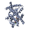
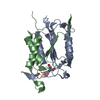
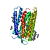
| Structure viewer | Molecule: MolmilJmol/JSmol |
|---|
- Downloads & links
-Download
| PDBx/mmCIF format | 1b8i.cif.gz | 58.5 KB | Display | PDBx/mmCIF format |
|---|---|---|---|---|
| PDB format | pdb1b8i.ent.gz | 40.4 KB | Display | PDB format |
| PDBx/mmJSON format | 1b8i.json.gz | Tree view | PDBx/mmJSON format | |
| Others |  Other downloads Other downloads |
-Validation report
| Arichive directory | https://data.pdbj.org/pub/pdb/validation_reports/b8/1b8iftp://data.pdbj.org/pub/pdb/validation_reports/b8/1b8i | HTTPS FTP |
|---|
-Related structure data
| Similar structure data |
|---|


-Links
 PDBj
PDBj


- Assembly
| Deposited unit | 
| ||||||||
|---|---|---|---|---|---|---|---|---|---|
| 1 |
| ||||||||
| Unit cell |
|
-Components
| #1: DNA chain | Mass: 4537.975 Da / Num. of mol.: 1 / Fragment: UBX/EXD CONSENSUS BINDING SITE / Source method: obtained synthetically |
|---|---|
| #2: DNA chain | Mass: 4640.020 Da / Num. of mol.: 1 / Fragment: UBX/EXD CONSENSUS BINDING SITE / Source method: obtained synthetically |
| #3: Protein | Mass: 9998.532 Da / Num. of mol.: 1 / Fragment: YPWM MOTIF AND HOMEODOMAIN / Mutation: C139S Source method: isolated from a genetically manipulated source Source: (gene. exp.) Drosophila melanogaster (fruit fly) / Description: IVA SPLICING ISOFORM OF THE UBX GENE WAS USED; / Cellular location: NUCLEUSCell nucleus / Gene: UBX / Plasmid: PET-3A / Cellular location (production host): CYTOPLASM / Production host:  Escherichia coli (E. coli) / Strain (production host): BL21(DE3) / Variant (production host): PLYSS / References: UniProt: P83949 Escherichia coli (E. coli) / Strain (production host): BL21(DE3) / Variant (production host): PLYSS / References: UniProt: P83949 |
| #4: Protein | Mass: 7553.561 Da / Num. of mol.: 1 / Fragment: HOMEODOMAIN Source method: isolated from a genetically manipulated source Source: (gene. exp.) Drosophila melanogaster (fruit fly) / Cellular location: NUCLEUSCell nucleus / Cellular location (production host): CYTOPLASM / Production host: Escherichia coli (E. coli) / References: UniProt: P40427 |
| #5: Water | ChemComp-HOH / Water Mass: 18.015 Da / Num. of mol.: 110 / Source method: isolated from a natural source / Formula: H2O Mass: 18.015 Da / Num. of mol.: 110 / Source method: isolated from a natural source / Formula: H2O |
-Experimental details
-Experiment
| Experiment | Method: X-RAY DIFFRACTION / Number of used crystals: 1 |
|---|
- Sample preparation
| Crystal | Density Matthews: 2.2 Å3/Da / Density % sol: 40 % |
|---|---|
| Crystal grow | Method: vapor diffusion, hanging drop / pH: 4.5 Details: 0.6M AMMONIUM PHOSPHATE, pH 4.5, VAPOR DIFFUSION, HANGING DROP |
| Crystal grow | *PLUS Method: unknown |
| Components of the solutions | *PLUS Conc.: 0.6 M / Common name: ammonium phosphate |
-Data collection
| Diffraction | Mean temperature: 113 K |
|---|---|
| Diffraction source | Source: SYNCHROTRON / Site: CHESS  / Beamline: F1 / Wavelength: 0.93 / Beamline: F1 / Wavelength: 0.93 |
| Detector | Type: ADSC / Detector: CCD / Date: Dec 15, 1998 |
| Radiation | Protocol: SINGLE WAVELENGTH / Monochromatic (M) / Laue (L): M / Scattering type: x-ray |
| Radiation wavelength | Wavelength: 0.93 Å / Relative weight: 1 |
| Reflection | Resolution: 2.4→20 Å / Num. obs: 10324 / % possible obs: 98.3 % / Redundancy: 10.5 % / Biso Wilson estimate: 48.2 Å2 / Rsym value: 7.4 |
| Reflection shell | Resolution: 2.4→2.49 Å / Rsym value: 18.5 / % possible all: 100 |
| Reflection | *PLUS Num. measured all: 110271 / Rmerge(I) obs: 0.074 |
| Reflection shell | *PLUS % possible obs: 100 % / Rmerge(I) obs: 0.185 |
- Processing
| Software |
| ||||||||||||||||||||||||||||||||||||||||||||||||||||||||||||||||||||||||||||||||
|---|---|---|---|---|---|---|---|---|---|---|---|---|---|---|---|---|---|---|---|---|---|---|---|---|---|---|---|---|---|---|---|---|---|---|---|---|---|---|---|---|---|---|---|---|---|---|---|---|---|---|---|---|---|---|---|---|---|---|---|---|---|---|---|---|---|---|---|---|---|---|---|---|---|---|---|---|---|---|---|---|---|
| Refinement | Method to determine structure: MIRAS / Resolution: 2.4→8 Å / Rfactor Rfree error: 0.014 / Data cutoff high absF: 10000000 / Data cutoff low absF: 0.001 / Isotropic thermal model: RESTRAINED / Cross valid method: THROUGHOUT / σ(F): 2 / Details: BULK SOLVENT MODEL USED
| ||||||||||||||||||||||||||||||||||||||||||||||||||||||||||||||||||||||||||||||||
| Displacement parameters | Biso mean: 42.7 Å2
| ||||||||||||||||||||||||||||||||||||||||||||||||||||||||||||||||||||||||||||||||
| Refine analyze |
| ||||||||||||||||||||||||||||||||||||||||||||||||||||||||||||||||||||||||||||||||
| Refinement step | Cycle: LAST / Resolution: 2.4→8 Å
| ||||||||||||||||||||||||||||||||||||||||||||||||||||||||||||||||||||||||||||||||
| Refine LS restraints |
| ||||||||||||||||||||||||||||||||||||||||||||||||||||||||||||||||||||||||||||||||
| LS refinement shell | Resolution: 2.4→2.51 Å / Rfactor Rfree error: 0.057 / Total num. of bins used: 8
| ||||||||||||||||||||||||||||||||||||||||||||||||||||||||||||||||||||||||||||||||
| Xplor file |
| ||||||||||||||||||||||||||||||||||||||||||||||||||||||||||||||||||||||||||||||||
| Software | *PLUS Name: X-PLOR / Version: 3.851 / Classification: refinement | ||||||||||||||||||||||||||||||||||||||||||||||||||||||||||||||||||||||||||||||||
| Refinement | *PLUS σ(F): 2 / % reflection Rfree: 5.4 % / Rfactor obs: 0.224 | ||||||||||||||||||||||||||||||||||||||||||||||||||||||||||||||||||||||||||||||||
| Solvent computation | *PLUS | ||||||||||||||||||||||||||||||||||||||||||||||||||||||||||||||||||||||||||||||||
| Displacement parameters | *PLUS Biso mean: 42.7 Å2 | ||||||||||||||||||||||||||||||||||||||||||||||||||||||||||||||||||||||||||||||||
| Refine LS restraints | *PLUS
| ||||||||||||||||||||||||||||||||||||||||||||||||||||||||||||||||||||||||||||||||
| LS refinement shell | *PLUS Rfactor Rfree: 0.324 / % reflection Rfree: 4.5 % / Rfactor Rwork: 0.32 |
