 Movie
Movie Controller

[English] 日本語
 Yorodumi
- PDB-5zby: Crystal structure of a [NiFe] hydrogenase maturation protease Hyc... -
+ Open data

- Basic information
| Entry | Database: PDB / ID: 5zby | |||||||||
|---|---|---|---|---|---|---|---|---|---|---|
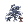
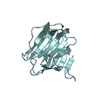
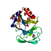
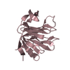
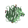
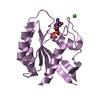
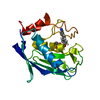
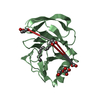
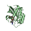
| Title | Crystal structure of a [NiFe] hydrogenase maturation protease HycI from Thermococcus kodakarensis KOD1 | |||||||||
 Components Components | Hydrogenase maturation protease HycI | |||||||||
 Keywords Keywords |  HYDROLASE / Maturation protease / HycI / [NiFe] hydrogenase / C-terminal cleavage HYDROLASE / Maturation protease / HycI / [NiFe] hydrogenase / C-terminal cleavage | |||||||||
| Function / homology | Peptidase A31, hydrogenase maturation protease HycI / Hydrogenase maturation protease / Peptidase A31 family / Peptidase HybD-like domain superfamily / protein modification process => GO:0036211 / enzyme activator activity / protein processing / endopeptidase activity / Hydrogenase maturation protease HycI Function and homology information Function and homology information | |||||||||
| Biological species |   Thermococcus kodakarensis KOD1 (archaea) Thermococcus kodakarensis KOD1 (archaea) | |||||||||
| Method | X-RAY DIFFRACTION / SYNCHROTRON / MOLECULAR REPLACEMENT / Resolution: 1.591 Å | |||||||||
 Authors Authors | Kwon, S. / Nishitani, Y. / Miki, K. | |||||||||
| Funding support |  Japan, 2items Japan, 2items
| |||||||||
 Citation Citation | Journal: Biochem. Biophys. Res. Commun. / Year: 2018 Title: Structure of a [NiFe] hydrogenase maturation protease HycI provides insights into its substrate selectivity Authors: Kwon, S. / Nishitani, Y. / Hirao, Y. / Kanai, T. / Atomi, H. / Miki, K. | |||||||||
| History |
|
- Structure visualization
| Structure viewer | Molecule: MolmilJmol/JSmol |
|---|
- Downloads & links
-Download
| PDBx/mmCIF format | 5zby.cif.gz | 45.6 KB | Display | PDBx/mmCIF format |
|---|---|---|---|---|
| PDB format | pdb5zby.ent.gz | 30.7 KB | Display | PDB format |
| PDBx/mmJSON format | 5zby.json.gz | Tree view | PDBx/mmJSON format | |
| Others |  Other downloads Other downloads |
-Validation report
| Arichive directory | https://data.pdbj.org/pub/pdb/validation_reports/zb/5zbyftp://data.pdbj.org/pub/pdb/validation_reports/zb/5zby | HTTPS FTP |
|---|
-Related structure data
| Related structure data |  2e85S S: Starting model for refinement |
|---|---|
| Similar structure data |

-Links
 PDBj
PDBj- Assembly
| Deposited unit | 
| ||||||||
|---|---|---|---|---|---|---|---|---|---|
| 1 |
| ||||||||
| Unit cell |
|
-Components
| #1: Protein | Mass: 17056.742 Da / Num. of mol.: 1 Source method: isolated from a genetically manipulated source Source: (gene. exp.) Thermococcus kodakarensis KOD1 (archaea)Strain: KOD1 / Gene: TK2004 / Production host:  Escherichia coli (E. coli) / References: UniProt: Q5JIH7 Escherichia coli (E. coli) / References: UniProt: Q5JIH7 |
|---|---|
| #2: Chemical | ChemComp-EPE / HEPES  Mass: 238.305 Da / Num. of mol.: 1 / Source method: obtained synthetically / Formula: C8H18N2O4S / Comment: pH buffer*YM Mass: 238.305 Da / Num. of mol.: 1 / Source method: obtained synthetically / Formula: C8H18N2O4S / Comment: pH buffer*YM |
| #3: Water | ChemComp-HOH / Water Mass: 18.015 Da / Num. of mol.: 102 / Source method: isolated from a natural source / Formula: H2O Mass: 18.015 Da / Num. of mol.: 102 / Source method: isolated from a natural source / Formula: H2O |
-Experimental details
-Experiment
| Experiment | Method: X-RAY DIFFRACTION / Number of used crystals: 1 |
|---|
- Sample preparation
| Crystal | Density Matthews: 2.72 Å3/Da / Density % sol: 54.75 % |
|---|---|
| Crystal grow | Temperature: 298 K / Method: vapor diffusion, hanging drop / Details: HEPES (pH 7.5), 2-methyl-2,4-pentanediol |
-Data collection
| Diffraction | Mean temperature: 95 K |
|---|---|
| Diffraction source | Source: SYNCHROTRON / Site: SPring-8 / Beamline: BL41XU / Wavelength: 1 Å |
| Detector | Type: DECTRIS PILATUS3 6M / Detector: PIXEL / Date: Oct 8, 2016 |
| Radiation | Protocol: SINGLE WAVELENGTH / Monochromatic (M) / Laue (L): M / Scattering type: x-ray |
| Radiation wavelength | Wavelength: 1 Å / Relative weight: 1 |
| Reflection | Resolution: 1.591→47.628 Å / Num. obs: 25807 / % possible obs: 98.1 % / Redundancy: 11.2 % / Rmerge(I) obs: 0.049 / Net I/σ(I): 35.4 |
| Reflection shell | Resolution: 1.591→1.63 Å / Rmerge(I) obs: 0.623 / Num. unique obs: 1300 |
- Processing
| Software |
| ||||||||||||||||||||||||||||||||||||||||||||||||||||||||||||||||||||||
|---|---|---|---|---|---|---|---|---|---|---|---|---|---|---|---|---|---|---|---|---|---|---|---|---|---|---|---|---|---|---|---|---|---|---|---|---|---|---|---|---|---|---|---|---|---|---|---|---|---|---|---|---|---|---|---|---|---|---|---|---|---|---|---|---|---|---|---|---|---|---|---|
| Refinement | Method to determine structure: MOLECULAR REPLACEMENT / Starting model: 2.0E+85 / Resolution: 1.591→47.628 Å / SU ML: 0.18 / Cross valid method: FREE R-VALUE / σ(F): 1.36 / Phase error: 28.41 / Stereochemistry target values: ML
| ||||||||||||||||||||||||||||||||||||||||||||||||||||||||||||||||||||||
| Solvent computation | Shrinkage radii: 0.9 Å / VDW probe radii: 1.11 Å / Solvent model: FLAT BULK SOLVENT MODEL | ||||||||||||||||||||||||||||||||||||||||||||||||||||||||||||||||||||||
| Refinement step | Cycle: LAST / Resolution: 1.591→47.628 Å
| ||||||||||||||||||||||||||||||||||||||||||||||||||||||||||||||||||||||
| Refine LS restraints |
| ||||||||||||||||||||||||||||||||||||||||||||||||||||||||||||||||||||||
| LS refinement shell |
|
