 Movie
Movie Controller

[English] 日本語
 Yorodumi
- PDB-5z75: Artificial L-threonine 3-dehydrogenase designed by ancestral sequ... -
+ Open data

- Basic information
| Entry | Database: PDB / ID: 5z75 | ||||||
|---|---|---|---|---|---|---|---|
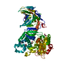
| Title | Artificial L-threonine 3-dehydrogenase designed by ancestral sequence reconstruction. | ||||||
 Components Components | Artificial L-threonine 3-dehydrogenase | ||||||
 Keywords Keywords |  OXIDOREDUCTASE / ancestral sequence reconstruction / L-threonine 3-dehydrogenase OXIDOREDUCTASE / ancestral sequence reconstruction / L-threonine 3-dehydrogenase | ||||||
| Function / homology | NAD(P)-binding Rossmann-like Domain / Rossmann fold / 3-Layer(aba) Sandwich / Alpha Beta / NICOTINAMIDE-ADENINE-DINUCLEOTIDE / PHOSPHATE ION Function and homology information Function and homology information | ||||||
| Biological species | synthetic construct (others) | ||||||
| Method | X-RAY DIFFRACTION / SYNCHROTRON / MOLECULAR REPLACEMENT / Resolution: 2.1 Å | ||||||
 Authors Authors | Nakano, S. / Motoyama, T. / Miyashita, Y. / Ishizuka, Y. / Matsuo, N. / Tokiwa, H. / Shinoda, S. / Asano, Y. / Ito, S. | ||||||
| Funding support |  Japan, 1items Japan, 1items
| ||||||
 Citation Citation | Journal: Biochemistry / Year: 2018 Title: Benchmark Analysis of Native and Artificial NAD+-Dependent Enzymes Generated by a Sequence-Based Design Method with or without Phylogenetic Data. Authors: Nakano, S. / Motoyama, T. / Miyashita, Y. / Ishizuka, Y. / Matsuo, N. / Tokiwa, H. / Shinoda, S. / Asano, Y. / Ito, S. | ||||||
| History |
|
- Structure visualization
| Structure viewer | Molecule: MolmilJmol/JSmol |
|---|
- Downloads & links
-Download
| PDBx/mmCIF format | 5z75.cif.gz | 269.4 KB | Display | PDBx/mmCIF format |
|---|---|---|---|---|
| PDB format | pdb5z75.ent.gz | 216.5 KB | Display | PDB format |
| PDBx/mmJSON format | 5z75.json.gz | Tree view | PDBx/mmJSON format | |
| Others |  Other downloads Other downloads |
-Validation report
| Arichive directory | https://data.pdbj.org/pub/pdb/validation_reports/z7/5z75ftp://data.pdbj.org/pub/pdb/validation_reports/z7/5z75 | HTTPS FTP |
|---|
-Related structure data
| Related structure data |  5z76C  3wmxS S: Starting model for refinement C: citing same article ( |
|---|---|
| Similar structure data |


-Links
 PDBj
PDBj- Assembly
| Deposited unit | 
| ||||||||||||||||||||||||||||||||||||||||||||||||||||||||||||||||||||||||||||||||||||||||||||||||||||||||||||||||||||||||||||
|---|---|---|---|---|---|---|---|---|---|---|---|---|---|---|---|---|---|---|---|---|---|---|---|---|---|---|---|---|---|---|---|---|---|---|---|---|---|---|---|---|---|---|---|---|---|---|---|---|---|---|---|---|---|---|---|---|---|---|---|---|---|---|---|---|---|---|---|---|---|---|---|---|---|---|---|---|---|---|---|---|---|---|---|---|---|---|---|---|---|---|---|---|---|---|---|---|---|---|---|---|---|---|---|---|---|---|---|---|---|---|---|---|---|---|---|---|---|---|---|---|---|---|---|---|---|
| 1 | 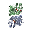
| ||||||||||||||||||||||||||||||||||||||||||||||||||||||||||||||||||||||||||||||||||||||||||||||||||||||||||||||||||||||||||||
| 2 | 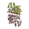
| ||||||||||||||||||||||||||||||||||||||||||||||||||||||||||||||||||||||||||||||||||||||||||||||||||||||||||||||||||||||||||||
| Unit cell |
| ||||||||||||||||||||||||||||||||||||||||||||||||||||||||||||||||||||||||||||||||||||||||||||||||||||||||||||||||||||||||||||
| Noncrystallographic symmetry (NCS) | NCS domain:
NCS domain segments: Component-ID: 0 / Beg auth comp-ID: MET / Beg label comp-ID: MET / End auth comp-ID: LEU / End label comp-ID: LEU / Refine code: 0
NCS ensembles :
|
-Components
| #1: Protein | Mass: 37009.844 Da / Num. of mol.: 4 Source method: isolated from a genetically manipulated source Source: (gene. exp.) synthetic construct (others) / Production host:  Escherichia coli (E. coli) Escherichia coli (E. coli)#2: Chemical | Nicotinamide adenine dinucleotide  Mass: 663.425 Da / Num. of mol.: 3 / Source method: obtained synthetically / Formula: C21H27N7O14P2 / Comment: NAD*YM Mass: 663.425 Da / Num. of mol.: 3 / Source method: obtained synthetically / Formula: C21H27N7O14P2 / Comment: NAD*YM#3: Chemical | ChemComp-PO4 / Phosphate  Mass: 94.971 Da / Num. of mol.: 7 / Source method: obtained synthetically / Formula: PO4 Mass: 94.971 Da / Num. of mol.: 7 / Source method: obtained synthetically / Formula: PO4#4: Chemical | Polyethylene glycol  Mass: 414.488 Da / Num. of mol.: 3 / Source method: obtained synthetically / Formula: C18H38O10 / Comment: precipitant*YM Mass: 414.488 Da / Num. of mol.: 3 / Source method: obtained synthetically / Formula: C18H38O10 / Comment: precipitant*YM#5: Water | ChemComp-HOH / | Water Mass: 18.015 Da / Num. of mol.: 522 / Source method: isolated from a natural source / Formula: H2O Mass: 18.015 Da / Num. of mol.: 522 / Source method: isolated from a natural source / Formula: H2O |
|---|
-Experimental details
-Experiment
| Experiment | Method: X-RAY DIFFRACTION / Number of used crystals: 1 |
|---|
- Sample preparation
| Crystal | Density Matthews: 2.17 Å3/Da / Density % sol: 43.37 % |
|---|---|
| Crystal grow | Temperature: 295 K / Method: vapor diffusion, sitting drop / Details: 20% PEG3350, 0.2M potassium phosphate monobasic |
-Data collection
| Diffraction | Mean temperature: 100 K |
|---|---|
| Diffraction source | Source: SYNCHROTRON / Site: SPring-8 / Beamline: BL38B1 / Wavelength: 1 Å |
| Detector | Type: MARMOSAIC 225 mm CCD / Detector: CCD / Date: Oct 13, 2017 |
| Radiation | Protocol: SINGLE WAVELENGTH / Monochromatic (M) / Laue (L): M / Scattering type: x-ray |
| Radiation wavelength | Wavelength: 1 Å / Relative weight: 1 |
| Reflection | Resolution: 2.1→92.7 Å / Num. obs: 73459 / % possible obs: 99.9 % / Redundancy: 7.4 % / Rmerge(I) obs: 0.087 / Net I/σ(I): 32.5 |
| Reflection shell | Resolution: 2.1→2.16 Å / Rmerge(I) obs: 0.428 / Mean I/σ(I) obs: 5.6 / Num. unique obs: 3652 / % possible all: 99.7 |
- Processing
| Software |
| ||||||||||||||||||||||||||||||||||||||||||||||||||||||||||||||||||||||||||||||||||||||||||||||||||||||||||||||||||||||||||||||||||||||||||||||||||||||||||||||||||||||||||||||||||||||
|---|---|---|---|---|---|---|---|---|---|---|---|---|---|---|---|---|---|---|---|---|---|---|---|---|---|---|---|---|---|---|---|---|---|---|---|---|---|---|---|---|---|---|---|---|---|---|---|---|---|---|---|---|---|---|---|---|---|---|---|---|---|---|---|---|---|---|---|---|---|---|---|---|---|---|---|---|---|---|---|---|---|---|---|---|---|---|---|---|---|---|---|---|---|---|---|---|---|---|---|---|---|---|---|---|---|---|---|---|---|---|---|---|---|---|---|---|---|---|---|---|---|---|---|---|---|---|---|---|---|---|---|---|---|---|---|---|---|---|---|---|---|---|---|---|---|---|---|---|---|---|---|---|---|---|---|---|---|---|---|---|---|---|---|---|---|---|---|---|---|---|---|---|---|---|---|---|---|---|---|---|---|---|---|
| Refinement | Method to determine structure: MOLECULAR REPLACEMENT Starting model: 3WMX Resolution: 2.1→92.7 Å / Cor.coef. Fo:Fc: 0.949 / Cor.coef. Fo:Fc free: 0.927 / SU B: 5.52 / SU ML: 0.145 / Cross valid method: THROUGHOUT / ESU R: 0.254 / ESU R Free: 0.195 / Details: HYDROGENS HAVE BEEN ADDED IN THE RIDING POSITIONS
| ||||||||||||||||||||||||||||||||||||||||||||||||||||||||||||||||||||||||||||||||||||||||||||||||||||||||||||||||||||||||||||||||||||||||||||||||||||||||||||||||||||||||||||||||||||||
| Solvent computation | Ion probe radii: 0.8 Å / Shrinkage radii: 0.8 Å / VDW probe radii: 1.2 Å | ||||||||||||||||||||||||||||||||||||||||||||||||||||||||||||||||||||||||||||||||||||||||||||||||||||||||||||||||||||||||||||||||||||||||||||||||||||||||||||||||||||||||||||||||||||||
| Displacement parameters | Biso mean: 36.065 Å2
| ||||||||||||||||||||||||||||||||||||||||||||||||||||||||||||||||||||||||||||||||||||||||||||||||||||||||||||||||||||||||||||||||||||||||||||||||||||||||||||||||||||||||||||||||||||||
| Refinement step | Cycle: 1 / Resolution: 2.1→92.7 Å
| ||||||||||||||||||||||||||||||||||||||||||||||||||||||||||||||||||||||||||||||||||||||||||||||||||||||||||||||||||||||||||||||||||||||||||||||||||||||||||||||||||||||||||||||||||||||
| Refine LS restraints |
|