 Movie
Movie Controller

[English] 日本語
 Yorodumi
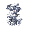
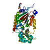
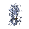
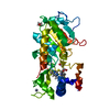
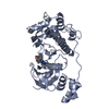
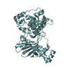
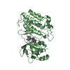
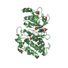
- PDB-5yql: Crystal structure of Sirt2 in complex with selective inhibitor A2I -
+ Open data

- Basic information
| Entry | Database: PDB / ID: 5yql | ||||||
|---|---|---|---|---|---|---|---|
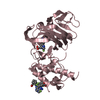
| Title | Crystal structure of Sirt2 in complex with selective inhibitor A2I | ||||||
 Components Components | NAD-dependent protein deacetylase sirtuin-2 | ||||||
 Keywords Keywords | HYDROLASE/HYDROLASE INHIBITOR / NAD-dependent deacetylase sirtuin-2 / Inhibitor /  Complex / HYDROLASE-HYDROLASE INHIBITOR complex Complex / HYDROLASE-HYDROLASE INHIBITOR complex | ||||||
| Function / homology |  Function and homology information Function and homology informationcellular response to caloric restriction / negative regulation of oligodendrocyte progenitor proliferation / cellular lipid catabolic process / negative regulation of striated muscle tissue development / : / NAD-dependent histone H4K16 deacetylase activity / positive regulation of meiotic nuclear division / positive regulation of attachment of spindle microtubules to kinetochore / NAD-dependent protein demyristoylase activity / NAD-dependent protein depalmitoylase activity ...cellular response to caloric restriction / negative regulation of oligodendrocyte progenitor proliferation / cellular lipid catabolic process / negative regulation of striated muscle tissue development / : / NAD-dependent histone H4K16 deacetylase activity / positive regulation of meiotic nuclear division / positive regulation of attachment of spindle microtubules to kinetochore / NAD-dependent protein demyristoylase activity / NAD-dependent protein depalmitoylase activity / paranodal junction / tubulin deacetylation / lateral loop / NLRP3 inflammasome complex assembly / peptidyl-lysine deacetylation / mitotic nuclear membrane reassembly / negative regulation of NLRP3 inflammasome complex assembly / tubulin deacetylase activity / regulation of exit from mitosis / paranode region of axon / Schmidt-Lanterman incisure / positive regulation of fatty acid biosynthetic process / NAD-dependent protein lysine deacetylase activity / myelination in peripheral nervous system / rDNA heterochromatin formation / protein acetyllysine N-acetyltransferase / chromatin silencing complex / NAD-dependent histone deacetylase activity / regulation of phosphorylation / Initiation of Nuclear Envelope (NE) Reformation / protein deacetylation / juxtaparanode region of axon / positive regulation of oocyte maturation / protein lysine deacetylase activity / meiotic spindle / response to redox state / regulation of myelination / histone deacetylase activity / histone acetyltransferase binding / positive regulation of execution phase of apoptosis / negative regulation of fat cell differentiation / negative regulation of peptidyl-threonine phosphorylation / glial cell projection / positive regulation of cell division / NAD+ ADP-ribosyltransferase activity / NAD+ binding / subtelomeric heterochromatin formation / negative regulation of reactive oxygen species metabolic process / positive regulation of DNA binding / heterochromatin / heterochromatin formation / epigenetic regulation of gene expression / Transferases; Acyltransferases; Transferring groups other than aminoacyl groups / cellular response to epinephrine stimulus / substantia nigra development / centriole / negative regulation of autophagy / meiotic cell cycle / ubiquitin binding / negative regulation of protein catabolic process / mitotic spindle / autophagy / histone deacetylase binding / spindle / positive regulation of proteasomal ubiquitin-dependent protein catabolic process / myelin sheath / chromosome / cellular response to oxidative stress / midbody / growth cone / cellular response to hypoxia / perikaryon / proteasome-mediated ubiquitin-dependent protein catabolic process / DNA-binding transcription factor binding / microtubule / chromosome, telomeric region / regulation of cell cycle / cell division / innate immune response / negative regulation of DNA-templated transcription / centrosome / chromatin binding / nucleolus / perinuclear region of cytoplasm / negative regulation of transcription by RNA polymerase II / positive regulation of transcription by RNA polymerase II / mitochondrion / zinc ion binding / nucleus / plasma membrane / cytosol / cytoplasmSimilarity search - Function | ||||||
| Biological species |  Homo sapiens (human) Homo sapiens (human) | ||||||
| Method | X-RAY DIFFRACTION / SYNCHROTRON / MOLECULAR REPLACEMENT / Resolution: 1.601 Å | ||||||
 Authors Authors | Wang, H. / Yu, Y. / Li, G. / Chen, Q. | ||||||
 Citation Citation | Journal: Eur J Med Chem / Year: 2018 Title: X-ray crystal structure guided discovery of new selective, substrate-mimicking sirtuin 2 inhibitors that exhibit activities against non-small cell lung cancer cells. Authors: Yang, L.L. / Wang, H.L. / Zhong, L. / Yuan, C. / Liu, S.Y. / Yu, Z.J. / Liu, S. / Yan, Y.H. / Wu, C. / Wang, Y. / Wang, Z. / Yu, Y. / Chen, Q. / Li, G.B. | ||||||
| History |
|
- Structure visualization
| Structure viewer | Molecule: MolmilJmol/JSmol |
|---|
- Downloads & links
-Download
| PDBx/mmCIF format | 5yql.cif.gz | 132.7 KB | Display | PDBx/mmCIF format |
|---|---|---|---|---|
| PDB format | pdb5yql.ent.gz | 100.9 KB | Display | PDB format |
| PDBx/mmJSON format | 5yql.json.gz | Tree view | PDBx/mmJSON format | |
| Others |  Other downloads Other downloads |
-Validation report
| Arichive directory | https://data.pdbj.org/pub/pdb/validation_reports/yq/5yqlftp://data.pdbj.org/pub/pdb/validation_reports/yq/5yql | HTTPS FTP |
|---|
-Related structure data
| Related structure data |  5yqmC  5yqnC  5yqoC  5matS S: Starting model for refinement C: citing same article ( |
|---|---|
| Similar structure data |

-Links
 PDBj
PDBj


- Assembly
| Deposited unit | 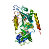
| ||||||||
|---|---|---|---|---|---|---|---|---|---|
| 1 |
| ||||||||
| Unit cell |
|
-Components
| #1: Protein | Mass: 34449.668 Da / Num. of mol.: 1 Source method: isolated from a genetically manipulated source Source: (gene. exp.) Homo sapiens (human) / Gene: SIRT2, SIR2L, SIR2L2 / Production host:  Escherichia coli (E. coli) Escherichia coli (E. coli)References: UniProt: Q8IXJ6, Hydrolases; Acting on carbon-nitrogen bonds, other than peptide bonds; In linear amides | ||
|---|---|---|---|
| #2: Chemical | ChemComp-A2I /   Mass: 379.475 Da / Num. of mol.: 1 / Source method: obtained synthetically / Formula: C21H21N3O2S / Feature type: SUBJECT OF INVESTIGATION Mass: 379.475 Da / Num. of mol.: 1 / Source method: obtained synthetically / Formula: C21H21N3O2S / Feature type: SUBJECT OF INVESTIGATION | ||
| #3: Chemical | ChemComp-ZN /   Mass: 65.409 Da / Num. of mol.: 1 / Source method: obtained synthetically / Formula: Zn Mass: 65.409 Da / Num. of mol.: 1 / Source method: obtained synthetically / Formula: Zn | ||
| #4: Chemical | 2-Mercaptoethanol  Mass: 78.133 Da / Num. of mol.: 2 / Source method: obtained synthetically / Formula: C2H6OS Mass: 78.133 Da / Num. of mol.: 2 / Source method: obtained synthetically / Formula: C2H6OS#5: Water | ChemComp-HOH / | Water Mass: 18.015 Da / Num. of mol.: 382 / Source method: isolated from a natural source / Formula: H2O Mass: 18.015 Da / Num. of mol.: 382 / Source method: isolated from a natural source / Formula: H2O |
-Experimental details
-Experiment
| Experiment | Method: X-RAY DIFFRACTION / Number of used crystals: 1 |
|---|
- Sample preparation
| Crystal | Density Matthews: 2.15 Å3/Da / Density % sol: 42.78 % |
|---|---|
| Crystal grow | Temperature: 293 K / Method: vapor diffusion, hanging drop Details: 0.2M Magnesium Formate, 23-30% (v/v) Polyethylene glycol 3350 |
-Data collection
| Diffraction | Mean temperature: 100 K |
|---|---|
| Diffraction source | Source: SYNCHROTRON / Site: SSRF  / Beamline: BL19U1 / Wavelength: 0.979 Å / Beamline: BL19U1 / Wavelength: 0.979 Å |
| Detector | Type: DECTRIS PILATUS 6M / Detector: PIXEL / Date: Feb 24, 2017 |
| Radiation | Protocol: SINGLE WAVELENGTH / Monochromatic (M) / Laue (L): M / Scattering type: x-ray |
| Radiation wavelength | Wavelength: 0.979 Å / Relative weight: 1 |
| Reflection | Resolution: 1.6→50 Å / Num. obs: 37723 / % possible obs: 98.2 % / Redundancy: 3.3 % / Rpim(I) all: 0.07 / Net I/σ(I): 10 |
| Reflection shell | Highest resolution: 1.6 Å / CC1/2: 0.791 / Rpim(I) all: 0.276 |
- Processing
| Software |
| |||||||||||||||||||||||||||||||||||||||||||||||||||||||||||||||
|---|---|---|---|---|---|---|---|---|---|---|---|---|---|---|---|---|---|---|---|---|---|---|---|---|---|---|---|---|---|---|---|---|---|---|---|---|---|---|---|---|---|---|---|---|---|---|---|---|---|---|---|---|---|---|---|---|---|---|---|---|---|---|---|---|
| Refinement | Method to determine structure: MOLECULAR REPLACEMENT Starting model: 5MAT Resolution: 1.601→35.895 Å / SU ML: 0.16 / Cross valid method: FREE R-VALUE / σ(F): 1.35 / Phase error: 19.46
| |||||||||||||||||||||||||||||||||||||||||||||||||||||||||||||||
| Solvent computation | Shrinkage radii: 0.9 Å / VDW probe radii: 1.11 Å | |||||||||||||||||||||||||||||||||||||||||||||||||||||||||||||||
| Refinement step | Cycle: LAST / Resolution: 1.601→35.895 Å
| |||||||||||||||||||||||||||||||||||||||||||||||||||||||||||||||
| Refine LS restraints |
| |||||||||||||||||||||||||||||||||||||||||||||||||||||||||||||||
| LS refinement shell |
|
