 Movie
Movie Controller


+ Open data

- Basic information
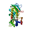
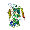
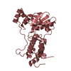
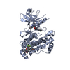
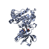
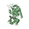
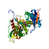
| Entry | Database: PDB / ID: 4rmh | ||||||
|---|---|---|---|---|---|---|---|
| Title | Human Sirt2 in complex with SirReal2 and Ac-Lys-H3 peptide | ||||||
 Components Components |
| ||||||
 Keywords Keywords | Hydrolase/Hydrolase Inhibitor / Hydrolase-Hydrolase Inhibitor complex | ||||||
| Function / homology |  Function and homology information Function and homology informationcellular response to caloric restriction / negative regulation of oligodendrocyte progenitor proliferation / cellular lipid catabolic process / negative regulation of striated muscle tissue development /  : / NAD-dependent histone H4K16 deacetylase activity / positive regulation of meiotic nuclear division / positive regulation of attachment of spindle microtubules to kinetochore / NAD-dependent protein demyristoylase activity / NAD-dependent protein depalmitoylase activity ...cellular response to caloric restriction / negative regulation of oligodendrocyte progenitor proliferation / cellular lipid catabolic process / negative regulation of striated muscle tissue development / : / NAD-dependent histone H4K16 deacetylase activity / positive regulation of meiotic nuclear division / positive regulation of attachment of spindle microtubules to kinetochore / NAD-dependent protein demyristoylase activity / NAD-dependent protein depalmitoylase activity / paranodal junction / tubulin deacetylation / lateral loop / NLRP3 inflammasome complex assembly / peptidyl-lysine deacetylation / mitotic nuclear membrane reassembly / negative regulation of NLRP3 inflammasome complex assembly / tubulin deacetylase activity / regulation of exit from mitosis / paranode region of axon / Schmidt-Lanterman incisure / positive regulation of fatty acid biosynthetic process / NAD-dependent protein lysine deacetylase activity / myelination in peripheral nervous system / rDNA heterochromatin formation / protein acetyllysine N-acetyltransferase / chromatin silencing complex / NAD-dependent histone deacetylase activity / regulation of phosphorylation / Initiation of Nuclear Envelope (NE) Reformation / protein deacetylation / juxtaparanode region of axon / positive regulation of oocyte maturation / protein lysine deacetylase activity / meiotic spindle / response to redox state / regulation of myelination / histone deacetylase activity / histone acetyltransferase binding / positive regulation of execution phase of apoptosis / negative regulation of fat cell differentiation / negative regulation of peptidyl-threonine phosphorylation / glial cell projection / positive regulation of cell division / NAD+ ADP-ribosyltransferase activity / NAD+ binding / subtelomeric heterochromatin formation / negative regulation of reactive oxygen species metabolic process / positive regulation of DNA binding / heterochromatin / heterochromatin formation / epigenetic regulation of gene expression / Transferases; Acyltransferases; Transferring groups other than aminoacyl groups / cellular response to epinephrine stimulus / substantia nigra development / centriole / negative regulation of autophagy / meiotic cell cycle / ubiquitin binding / negative regulation of protein catabolic process / mitotic spindle / autophagy / histone deacetylase binding / spindle / positive regulation of proteasomal ubiquitin-dependent protein catabolic process / myelin sheath / chromosome / cellular response to oxidative stress / midbody / growth cone / cellular response to hypoxia / perikaryon / proteasome-mediated ubiquitin-dependent protein catabolic process / DNA-binding transcription factor binding / microtubule / chromosome, telomeric region / regulation of cell cycle / cell division / innate immune response / negative regulation of DNA-templated transcription / centrosome / chromatin binding / nucleolus / perinuclear region of cytoplasm / negative regulation of transcription by RNA polymerase II / positive regulation of transcription by RNA polymerase II / mitochondrion / zinc ion binding / nucleus / plasma membrane / cytosol / cytoplasm : / NAD-dependent histone H4K16 deacetylase activity / positive regulation of meiotic nuclear division / positive regulation of attachment of spindle microtubules to kinetochore / NAD-dependent protein demyristoylase activity / NAD-dependent protein depalmitoylase activity ...cellular response to caloric restriction / negative regulation of oligodendrocyte progenitor proliferation / cellular lipid catabolic process / negative regulation of striated muscle tissue development / : / NAD-dependent histone H4K16 deacetylase activity / positive regulation of meiotic nuclear division / positive regulation of attachment of spindle microtubules to kinetochore / NAD-dependent protein demyristoylase activity / NAD-dependent protein depalmitoylase activity / paranodal junction / tubulin deacetylation / lateral loop / NLRP3 inflammasome complex assembly / peptidyl-lysine deacetylation / mitotic nuclear membrane reassembly / negative regulation of NLRP3 inflammasome complex assembly / tubulin deacetylase activity / regulation of exit from mitosis / paranode region of axon / Schmidt-Lanterman incisure / positive regulation of fatty acid biosynthetic process / NAD-dependent protein lysine deacetylase activity / myelination in peripheral nervous system / rDNA heterochromatin formation / protein acetyllysine N-acetyltransferase / chromatin silencing complex / NAD-dependent histone deacetylase activity / regulation of phosphorylation / Initiation of Nuclear Envelope (NE) Reformation / protein deacetylation / juxtaparanode region of axon / positive regulation of oocyte maturation / protein lysine deacetylase activity / meiotic spindle / response to redox state / regulation of myelination / histone deacetylase activity / histone acetyltransferase binding / positive regulation of execution phase of apoptosis / negative regulation of fat cell differentiation / negative regulation of peptidyl-threonine phosphorylation / glial cell projection / positive regulation of cell division / NAD+ ADP-ribosyltransferase activity / NAD+ binding / subtelomeric heterochromatin formation / negative regulation of reactive oxygen species metabolic process / positive regulation of DNA binding / heterochromatin / heterochromatin formation / epigenetic regulation of gene expression / Transferases; Acyltransferases; Transferring groups other than aminoacyl groups / cellular response to epinephrine stimulus / substantia nigra development / centriole / negative regulation of autophagy / meiotic cell cycle / ubiquitin binding / negative regulation of protein catabolic process / mitotic spindle / autophagy / histone deacetylase binding / spindle / positive regulation of proteasomal ubiquitin-dependent protein catabolic process / myelin sheath / chromosome / cellular response to oxidative stress / midbody / growth cone / cellular response to hypoxia / perikaryon / proteasome-mediated ubiquitin-dependent protein catabolic process / DNA-binding transcription factor binding / microtubule / chromosome, telomeric region / regulation of cell cycle / cell division / innate immune response / negative regulation of DNA-templated transcription / centrosome / chromatin binding / nucleolus / perinuclear region of cytoplasm / negative regulation of transcription by RNA polymerase II / positive regulation of transcription by RNA polymerase II / mitochondrion / zinc ion binding / nucleus / plasma membrane / cytosol / cytoplasmSimilarity search - Function | ||||||
| Biological species |  Homo sapiens (human) Homo sapiens (human) | ||||||
| Method | X-RAY DIFFRACTION / SYNCHROTRON / MOLECULAR REPLACEMENT / Resolution: 1.42 Å | ||||||
 Authors Authors | Rumpf, T. / Schiedel, M. / Karaman, B. / Roessler, C. / North, B.J. / Lehotzky, A. / Olah, J. / Ladwein, K.I. / Schmidtkunz, K. / Gajer, M. ...Rumpf, T. / Schiedel, M. / Karaman, B. / Roessler, C. / North, B.J. / Lehotzky, A. / Olah, J. / Ladwein, K.I. / Schmidtkunz, K. / Gajer, M. / Pannek, M. / Steegborn, C. / Sinclair, D.A. / Gerhardt, S. / Ovadi, J. / Schutkowski, M. / Sippl, W. / Einsle, O. / Jung, M. | ||||||
 Citation Citation | Journal: Nat Commun / Year: 2015 Title: Selective Sirt2 inhibition by ligand-induced rearrangement of the active site. Authors: Rumpf, T. / Schiedel, M. / Karaman, B. / Roessler, C. / North, B.J. / Lehotzky, A. / Olah, J. / Ladwein, K.I. / Schmidtkunz, K. / Gajer, M. / Pannek, M. / Steegborn, C. / Sinclair, D.A. / ...Authors: Rumpf, T. / Schiedel, M. / Karaman, B. / Roessler, C. / North, B.J. / Lehotzky, A. / Olah, J. / Ladwein, K.I. / Schmidtkunz, K. / Gajer, M. / Pannek, M. / Steegborn, C. / Sinclair, D.A. / Gerhardt, S. / Ovadi, J. / Schutkowski, M. / Sippl, W. / Einsle, O. / Jung, M. | ||||||
| History |
|
- Structure visualization
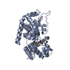
| Structure viewer | Molecule: MolmilJmol/JSmol |
|---|
- Downloads & links
-Download
| PDBx/mmCIF format | 4rmh.cif.gz | 134.7 KB | Display | PDBx/mmCIF format |
|---|---|---|---|---|
| PDB format | pdb4rmh.ent.gz | 103.6 KB | Display | PDB format |
| PDBx/mmJSON format | 4rmh.json.gz | Tree view | PDBx/mmJSON format | |
| Others |  Other downloads Other downloads |
-Validation report
| Arichive directory | https://data.pdbj.org/pub/pdb/validation_reports/rm/4rmhftp://data.pdbj.org/pub/pdb/validation_reports/rm/4rmh | HTTPS FTP |
|---|
-Related structure data


-Links
 PDBj
PDBj


- Assembly
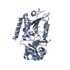
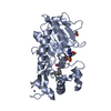
| Deposited unit | 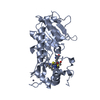
| ||||||||
|---|---|---|---|---|---|---|---|---|---|
| 1 |
| ||||||||
| Unit cell |
|
-Components
| #1: Protein | Mass: 34416.727 Da / Num. of mol.: 1 / Fragment: UNP RESIDUES 56-356 Source method: isolated from a genetically manipulated source Source: (gene. exp.) Homo sapiens (human) / Gene: SIRT2, SIR2L, SIR2L2 / Production host:  Escherichia coli (E. coli) Escherichia coli (E. coli)References: UniProt: Q8IXJ6, Hydrolases; Acting on carbon-nitrogen bonds, other than peptide bonds; In linear amides |
|---|---|
| #2: Protein/peptide | Mass: 728.818 Da / Num. of mol.: 1 / Source method: obtained synthetically |
| #3: Chemical | ChemComp-ZN /   Mass: 65.409 Da / Num. of mol.: 1 / Source method: obtained synthetically / Formula: Zn Mass: 65.409 Da / Num. of mol.: 1 / Source method: obtained synthetically / Formula: Zn |
| #4: Chemical | ChemComp-3TE /   Mass: 420.550 Da / Num. of mol.: 1 / Source method: obtained synthetically / Formula: C22H20N4OS2 Mass: 420.550 Da / Num. of mol.: 1 / Source method: obtained synthetically / Formula: C22H20N4OS2 |
| #5: Water | ChemComp-HOH / Water Mass: 18.015 Da / Num. of mol.: 243 / Source method: isolated from a natural source / Formula: H2O Mass: 18.015 Da / Num. of mol.: 243 / Source method: isolated from a natural source / Formula: H2O |
-Experimental details
-Experiment
| Experiment | Method: X-RAY DIFFRACTION / Number of used crystals: 1 |
|---|
- Sample preparation
| Crystal | Density Matthews: 2.07 Å3/Da / Density % sol: 40.49 % |
|---|---|
| Crystal grow | Temperature: 277 K / Method: vapor diffusion, sitting drop / pH: 9 Details: 2.8 M ammonium sulfate, pH 9.0, VAPOR DIFFUSION, SITTING DROP, temperature 277K |
-Data collection
| Diffraction | Mean temperature: 100 K |
|---|---|
| Diffraction source | Source: SYNCHROTRON / Site: SLS  / Beamline: X06SA / Wavelength: 1 Å / Beamline: X06SA / Wavelength: 1 Å |
| Detector | Type: DECTRIS PILATUS 6M / Detector: PIXEL / Date: May 8, 2013 |
| Radiation | Monochromator: FIXED-EXIT LN2 COOLED DOUBLE CRYSTAL MONOCHROMATOR Protocol: SINGLE WAVELENGTH / Monochromatic (M) / Laue (L): M / Scattering type: x-ray |
| Radiation wavelength | Wavelength: 1 Å / Relative weight: 1 |
| Reflection | Resolution: 1.42→55.06 Å / Num. obs: 53705 / % possible obs: 99.6 % / Observed criterion σ(F): 0 / Observed criterion σ(I): 1.5 |
| Reflection shell | Resolution: 1.42→1.44 Å / % possible all: 99.6 |
- Processing
| Software |
| ||||||||||||||||||||||||||||||||||||||||||||||||||||||||||||||||||||||||||||||||||||||||||||||||||||||||||||||||||||||||||||||||||||||||||||||||||||||||||||||||||||||||||||||||||||||
|---|---|---|---|---|---|---|---|---|---|---|---|---|---|---|---|---|---|---|---|---|---|---|---|---|---|---|---|---|---|---|---|---|---|---|---|---|---|---|---|---|---|---|---|---|---|---|---|---|---|---|---|---|---|---|---|---|---|---|---|---|---|---|---|---|---|---|---|---|---|---|---|---|---|---|---|---|---|---|---|---|---|---|---|---|---|---|---|---|---|---|---|---|---|---|---|---|---|---|---|---|---|---|---|---|---|---|---|---|---|---|---|---|---|---|---|---|---|---|---|---|---|---|---|---|---|---|---|---|---|---|---|---|---|---|---|---|---|---|---|---|---|---|---|---|---|---|---|---|---|---|---|---|---|---|---|---|---|---|---|---|---|---|---|---|---|---|---|---|---|---|---|---|---|---|---|---|---|---|---|---|---|---|---|
| Refinement | Method to determine structure: MOLECULAR REPLACEMENT / Resolution: 1.42→44.02 Å / Cor.coef. Fo:Fc: 0.966 / Cor.coef. Fo:Fc free: 0.966 / SU B: 2.546 / SU ML: 0.049 / Cross valid method: THROUGHOUT / σ(F): 0 / ESU R: 0.068 / ESU R Free: 0.062 / Stereochemistry target values: MAXIMUM LIKELIHOOD / Details: HYDROGENS HAVE BEEN ADDED IN THE RIDING POSITIONS
| ||||||||||||||||||||||||||||||||||||||||||||||||||||||||||||||||||||||||||||||||||||||||||||||||||||||||||||||||||||||||||||||||||||||||||||||||||||||||||||||||||||||||||||||||||||||
| Solvent computation | Ion probe radii: 0.8 Å / Shrinkage radii: 0.8 Å / VDW probe radii: 1.2 Å / Solvent model: MASK | ||||||||||||||||||||||||||||||||||||||||||||||||||||||||||||||||||||||||||||||||||||||||||||||||||||||||||||||||||||||||||||||||||||||||||||||||||||||||||||||||||||||||||||||||||||||
| Displacement parameters | Biso mean: 21.173 Å2
| ||||||||||||||||||||||||||||||||||||||||||||||||||||||||||||||||||||||||||||||||||||||||||||||||||||||||||||||||||||||||||||||||||||||||||||||||||||||||||||||||||||||||||||||||||||||
| Refinement step | Cycle: LAST / Resolution: 1.42→44.02 Å
| ||||||||||||||||||||||||||||||||||||||||||||||||||||||||||||||||||||||||||||||||||||||||||||||||||||||||||||||||||||||||||||||||||||||||||||||||||||||||||||||||||||||||||||||||||||||
| Refine LS restraints |
|