 Movie
Movie Controller

[English] 日本語
 Yorodumi
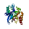
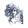
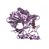
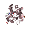
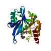
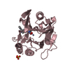
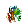
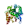
- PDB-5ypl: Crystal structure of NDM-1 bound to hydrolyzed imipenem represent... -
+ Open data

- Basic information
| Entry | Database: PDB / ID: 5ypl | ||||||
|---|---|---|---|---|---|---|---|
| Title | Crystal structure of NDM-1 bound to hydrolyzed imipenem representing an EP complex | ||||||
 Components Components | Metallo-beta-lactamase NDM-1 | ||||||
 Keywords Keywords |  HYDROLASE / NDM-1 / Imipenem / EP complex HYDROLASE / NDM-1 / Imipenem / EP complex | ||||||
| Function / homology |  Function and homology information Function and homology informationantibiotic catabolic process / beta-lactamase / hydrolase activity / metal ion bindingSimilarity search - Function | ||||||
| Biological species |  Escherichia coli (E. coli) Escherichia coli (E. coli) | ||||||
| Method | X-RAY DIFFRACTION / SYNCHROTRON / MOLECULAR REPLACEMENT / Resolution: 1.8 Å | ||||||
 Authors Authors | Feng, H. / Wang, D. / Liu, W. | ||||||
 Citation Citation | Journal: Nat Commun / Year: 2017 Title: The mechanism of NDM-1-catalyzed carbapenem hydrolysis is distinct from that of penicillin or cephalosporin hydrolysis. Authors: Feng, H. / Liu, X. / Wang, S. / Fleming, J. / Wang, D.C. / Liu, W. | ||||||
| History |
|
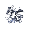
- Structure visualization
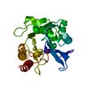
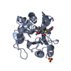
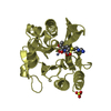
| Structure viewer | Molecule: MolmilJmol/JSmol |
|---|
- Downloads & links
-Download
| PDBx/mmCIF format | 5ypl.cif.gz | 191.9 KB | Display | PDBx/mmCIF format |
|---|---|---|---|---|
| PDB format | pdb5ypl.ent.gz | 150.8 KB | Display | PDB format |
| PDBx/mmJSON format | 5ypl.json.gz | Tree view | PDBx/mmJSON format | |
| Others |  Other downloads Other downloads |
-Validation report
| Arichive directory | https://data.pdbj.org/pub/pdb/validation_reports/yp/5yplftp://data.pdbj.org/pub/pdb/validation_reports/yp/5ypl | HTTPS FTP |
|---|
-Related structure data
| Related structure data |  5ypiC  5ypkC  5ypmC  5ypnC  4rl0S S: Starting model for refinement C: citing same article ( |
|---|---|
| Similar structure data |


-Links
 PDBj
PDBj


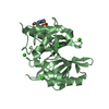
- Assembly
| Deposited unit | 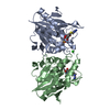
| ||||||||
|---|---|---|---|---|---|---|---|---|---|
| 1 | 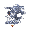
| ||||||||
| 2 | 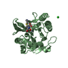
| ||||||||
| Unit cell |
|
-Components
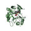
-Protein , 1 types, 2 molecules AB
| #1: Protein | Mass: 25631.820 Da / Num. of mol.: 2 Source method: isolated from a genetically manipulated source Source: (gene. exp.) Escherichia coli (E. coli) / Gene: blaNDM-1 / Production host: Escherichia coli (E. coli) / References: UniProt: A0A0A7Y424, UniProt: E5KIY2*PLUS |
|---|
-Non-polymers , 5 types, 379 molecules 
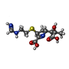


| #2: Chemical | ChemComp-ZN /  Mass: 65.409 Da / Num. of mol.: 4 / Source method: obtained synthetically / Formula: Zn Mass: 65.409 Da / Num. of mol.: 4 / Source method: obtained synthetically / Formula: Zn#3: Chemical |  Mass: 317.361 Da / Num. of mol.: 2 / Source method: obtained synthetically / Formula: C12H19N3O5S Mass: 317.361 Da / Num. of mol.: 2 / Source method: obtained synthetically / Formula: C12H19N3O5S#4: Chemical | ChemComp-SO4 / | Sulfate Mass: 96.063 Da / Num. of mol.: 1 / Source method: obtained synthetically / Formula: SO4 Mass: 96.063 Da / Num. of mol.: 1 / Source method: obtained synthetically / Formula: SO4#5: Chemical | ChemComp-CL / | Chloride Mass: 35.453 Da / Num. of mol.: 1 / Source method: obtained synthetically / Formula: Cl Mass: 35.453 Da / Num. of mol.: 1 / Source method: obtained synthetically / Formula: Cl#6: Water | ChemComp-HOH / | WaterMass: 18.015 Da / Num. of mol.: 371 / Source method: isolated from a natural source / Formula: H2O |
|---|
-Experimental details
-Experiment
| Experiment | Method: X-RAY DIFFRACTION / Number of used crystals: 1 |
|---|
- Sample preparation
| Crystal | Density Matthews: 1.93 Å3/Da / Density % sol: 36.2 % |
|---|---|
| Crystal grow | Temperature: 295 K / Method: vapor diffusion, hanging drop Details: 28% (w/v) PEG3350, 0.1M Bis-Tris, pH 5.8, 0.2M ammonium sulfate |
-Data collection
| Diffraction | Mean temperature: 100 K |
|---|---|
| Diffraction source | Source: SYNCHROTRON / Site: SSRF  / Beamline: BL19U1 / Wavelength: 0.97845 Å / Beamline: BL19U1 / Wavelength: 0.97845 Å |
| Detector | Type: DECTRIS PILATUS 6M / Detector: PIXEL / Date: May 24, 2015 |
| Radiation | Protocol: SINGLE WAVELENGTH / Monochromatic (M) / Laue (L): M / Scattering type: x-ray |
| Radiation wavelength | Wavelength: 0.97845 Å / Relative weight: 1 |
| Reflection | Resolution: 1.8→42.28 Å / Num. obs: 35229 / % possible obs: 94 % / Redundancy: 6.36 % / Rsym value: 0.07 / Net I/σ(I): 16.8 |
| Reflection shell | Resolution: 1.8→1.84 Å / Redundancy: 4.44 % / Mean I/σ(I) obs: 2.55 / Rsym value: 0.555 / % possible all: 64.8 |
- Processing
| Software |
| ||||||||||||||||
|---|---|---|---|---|---|---|---|---|---|---|---|---|---|---|---|---|---|
| Refinement | Method to determine structure: MOLECULAR REPLACEMENT Starting model: 4RL0 Resolution: 1.8→42.28 Å / SU ML: 0.17 / Cross valid method: FREE R-VALUE / σ(F): 1.36 / Phase error: 20.73 Details: THE STRUCTURE FACTOR FILE CONTAINS FRIEDEL PAIRS IN F_PLUS/MINUS COLUMNS
| ||||||||||||||||
| Solvent computation | Shrinkage radii: 0.9 Å / VDW probe radii: 1.11 Å | ||||||||||||||||
| Refinement step | Cycle: LAST / Resolution: 1.8→42.28 Å
|
