 Movie
Movie Controller

[English] 日本語
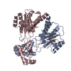
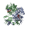
 Yorodumi
- PDB-5yii: Crystal Structure of 45 amino acid deleted from N-terminal of Pho... -
+ Open data

- Basic information
| Entry | Database: PDB / ID: 5yii | ||||||
|---|---|---|---|---|---|---|---|
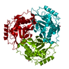
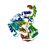
| Title | Crystal Structure of 45 amino acid deleted from N-terminal of Phosphoserine Aminotransferase (PSAT) of Entamoeba histolytica | ||||||
 Components Components | Phosphoserine aminotransferase Phosphoserine transaminase Phosphoserine transaminase | ||||||
 Keywords Keywords | TRANSFERASE / Delta45_EhPSAT | ||||||
| Function / homology |  Function and homology informationphosphoserine transaminase / O-phospho-L-serine:2-oxoglutarate aminotransferase activity / L-serine biosynthetic process / pyridoxal phosphate binding / cytoplasm Function and homology informationphosphoserine transaminase / O-phospho-L-serine:2-oxoglutarate aminotransferase activity / L-serine biosynthetic process / pyridoxal phosphate binding / cytoplasmSimilarity search - Function | ||||||
| Biological species |   Entamoeba histolytica (eukaryote) Entamoeba histolytica (eukaryote) | ||||||
| Method | X-RAY DIFFRACTION / SYNCHROTRON / MOLECULAR REPLACEMENT / molecular replacement / Resolution: 1.8 Å | ||||||
 Authors Authors | Singh, R.K. / Gourinath, S. | ||||||
 Citation Citation | Journal: Int.J.Biol.Macromol. / Year: 2019 Title: N-terminal residues are crucial for quaternary structure and active site conformation for the phosphoserine aminotransferase from enteric human parasite E. histolytica. Authors: Singh, R.K. / Tomar, P. / Dharavath, S. / Kumar, S. / Gourinath, S. | ||||||
| History |
|
- Structure visualization
| Structure viewer | Molecule: MolmilJmol/JSmol |
|---|
- Downloads & links
-Download
| PDBx/mmCIF format | 5yii.cif.gz | 77.6 KB | Display | PDBx/mmCIF format |
|---|---|---|---|---|
| PDB format | pdb5yii.ent.gz | 56.6 KB | Display | PDB format |
| PDBx/mmJSON format | 5yii.json.gz | Tree view | PDBx/mmJSON format | |
| Others |  Other downloads Other downloads |
-Validation report
| Arichive directory | https://data.pdbj.org/pub/pdb/validation_reports/yi/5yiiftp://data.pdbj.org/pub/pdb/validation_reports/yi/5yii | HTTPS FTP |
|---|
-Related structure data
| Related structure data | 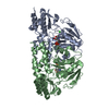 5yb0SC  5yd2C S: Starting model for refinement C: citing same article ( |
|---|---|
| Similar structure data |


-Links
 PDBj
PDBj
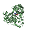
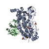
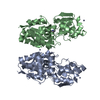
- Assembly
| Deposited unit | 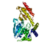
| ||||||||
|---|---|---|---|---|---|---|---|---|---|
| 1 | 
| ||||||||
| Unit cell |
|
-Components
| #1: Protein | Phosphoserine transaminase / Phosphoserine aminotransferase / putative Mass: 35628.328 Da / Num. of mol.: 1 Source method: isolated from a genetically manipulated source Source: (gene. exp.) Entamoeba histolytica (eukaryote) / Gene: EhPSAT, EHI_026360 / Plasmid: pET21C / Production host:  Escherichia coli (E. coli) / References: UniProt: Q60I38, phosphoserine transaminase Escherichia coli (E. coli) / References: UniProt: Q60I38, phosphoserine transaminase |
|---|---|
| #2: Chemical | ChemComp-SCN / Thiocyanate  Mass: 58.082 Da / Num. of mol.: 1 / Source method: obtained synthetically / Formula: CNS Mass: 58.082 Da / Num. of mol.: 1 / Source method: obtained synthetically / Formula: CNS |
| #3: Water | ChemComp-HOH / Water Mass: 18.015 Da / Num. of mol.: 87 / Source method: isolated from a natural source / Formula: H2O Mass: 18.015 Da / Num. of mol.: 87 / Source method: isolated from a natural source / Formula: H2O |
-Experimental details
-Experiment
| Experiment | Method: X-RAY DIFFRACTION / Number of used crystals: 1 |
|---|
- Sample preparation
| Crystal | Density Matthews: 2.13 Å3/Da / Density % sol: 42.18 % Description: The entry contains friedel pairs in F_plus/minus columns and I_plus/minus columns |
|---|---|
| Crystal grow | Temperature: 277.15 K / Method: vapor diffusion, sitting drop Details: 25% PEG 2000MME, 100 mM potassium thiocyanate (KSCN) |
-Data collection
| Diffraction | Mean temperature: 77.15 K |
|---|---|
| Diffraction source | Source: SYNCHROTRON / Site: ESRF  / Beamline: BM14 / Wavelength: 0.97 Å / Beamline: BM14 / Wavelength: 0.97 Å |
| Detector | Type: MAR CCD 165 mm / Detector: CCD / Date: Nov 30, 2015 |
| Radiation | Monochromator: Si 111 CHANNEL / Protocol: SINGLE WAVELENGTH / Monochromatic (M) / Laue (L): M / Scattering type: x-ray |
| Radiation wavelength | Wavelength: 0.97 Å / Relative weight: 1 |
| Reflection | Resolution: 1.8→50 Å / Num. obs: 54165 / % possible obs: 99.9 % / Redundancy: 3.8 % / Net I/σ(I): 41.93 |
| Reflection shell | Resolution: 1.8→1.83 Å |
-Phasing
| Phasing | Method: molecular replacement |
|---|
- Processing
| Software |
| ||||||||||||||||||||||||||||||||||||||||||||||||||||||||||||
|---|---|---|---|---|---|---|---|---|---|---|---|---|---|---|---|---|---|---|---|---|---|---|---|---|---|---|---|---|---|---|---|---|---|---|---|---|---|---|---|---|---|---|---|---|---|---|---|---|---|---|---|---|---|---|---|---|---|---|---|---|---|
| Refinement | Method to determine structure: MOLECULAR REPLACEMENT Starting model: 5YB0 Resolution: 1.8→50 Å / Cor.coef. Fo:Fc: 0.95 / Cor.coef. Fo:Fc free: 0.937 / SU B: 4.51 / SU ML: 0.137 / Cross valid method: THROUGHOUT / σ(F): 0 / ESU R: 0.167 / ESU R Free: 0.154 Details: The entry contains friedel pairs in F_plus/minus columns and I_plus/minus columns. HYDROGENS HAVE BEEN ADDED IN THE RIDING POSITIONS U VALUES : REFINED INDIVIDUALLY
| ||||||||||||||||||||||||||||||||||||||||||||||||||||||||||||
| Solvent computation | Ion probe radii: 0.8 Å / Shrinkage radii: 0.8 Å / VDW probe radii: 1.2 Å | ||||||||||||||||||||||||||||||||||||||||||||||||||||||||||||
| Displacement parameters | Biso max: 231.05 Å2 / Biso mean: 38.607 Å2 / Biso min: 19.32 Å2
| ||||||||||||||||||||||||||||||||||||||||||||||||||||||||||||
| Refinement step | Cycle: final / Resolution: 1.8→50 Å
| ||||||||||||||||||||||||||||||||||||||||||||||||||||||||||||
| Refine LS restraints |
| ||||||||||||||||||||||||||||||||||||||||||||||||||||||||||||
| LS refinement shell | Resolution: 1.8→1.847 Å / Rfactor Rfree error: 0 / Total num. of bins used: 20
|
