 Movie
Movie Controller

[English] 日本語
 Yorodumi
- PDB-5ygc: Crystal structure of Drosophila melanogaster Papi extended Tudor ... -
+ Open data

- Basic information
| Entry | Database: PDB / ID: 5ygc | ||||||
|---|---|---|---|---|---|---|---|
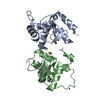
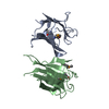
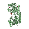
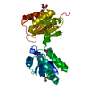
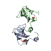
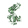
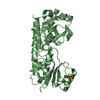
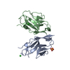
| Title | Crystal structure of Drosophila melanogaster Papi extended Tudor domain | ||||||
 Components Components | GH18329p | ||||||
 Keywords Keywords |  PROTEIN BINDING / piRNA / Papi / extended Tudor domain PROTEIN BINDING / piRNA / Papi / extended Tudor domain | ||||||
| Function / homology |  Function and homology information Function and homology informationprimary piRNA processing / P granule organization / piRNA processing / retrotransposon silencing / P granule / P-body / spermatogenesis / protein-containing complex / mitochondrion / RNA binding / nucleusSimilarity search - Function | ||||||
| Biological species |  Drosophila melanogaster (fruit fly) Drosophila melanogaster (fruit fly) | ||||||
| Method | X-RAY DIFFRACTION / SYNCHROTRON / MOLECULAR REPLACEMENT / Resolution: 2 Å | ||||||
 Authors Authors | Zhang, Y.H. / Huang, Y. | ||||||
 Citation Citation | Journal: Proc. Natl. Acad. Sci. U.S.A. / Year: 2018 Title: Structural insights into the sequence-specific recognition of Piwi byDrosophilaPapi Authors: Zhang, Y. / Liu, W. / Li, R. / Gu, J. / Wu, P. / Peng, C. / Ma, J. / Wu, L. / Yu, Y. / Huang, Y. | ||||||
| History |
|
- Structure visualization
| Structure viewer | Molecule: MolmilJmol/JSmol |
|---|
- Downloads & links
-Download
| PDBx/mmCIF format | 5ygc.cif.gz | 60.7 KB | Display | PDBx/mmCIF format |
|---|---|---|---|---|
| PDB format | pdb5ygc.ent.gz | 42.4 KB | Display | PDB format |
| PDBx/mmJSON format | 5ygc.json.gz | Tree view | PDBx/mmJSON format | |
| Others |  Other downloads Other downloads |
-Validation report
| Arichive directory | https://data.pdbj.org/pub/pdb/validation_reports/yg/5ygcftp://data.pdbj.org/pub/pdb/validation_reports/yg/5ygc | HTTPS FTP |
|---|
-Related structure data
| Related structure data | 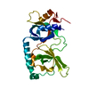 5ygbC  5ygdC  5ygfC  3fdrS S: Starting model for refinement C: citing same article ( |
|---|---|
| Similar structure data |


-Links
 PDBj
PDBj
- Assembly
| Deposited unit | 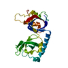
| |||||||||
|---|---|---|---|---|---|---|---|---|---|---|
| 1 |
| |||||||||
| Unit cell |
| |||||||||
| Components on special symmetry positions |
|
-Components
| #1: Protein | Mass: 25156.574 Da / Num. of mol.: 1 / Fragment: UNP residues 259-479 Source method: isolated from a genetically manipulated source Source: (gene. exp.) Drosophila melanogaster (fruit fly) / Gene: papi, Dmel\CG7082, PAPI, Papi, CG7082, Dmel_CG7082 / Production host:  Escherichia coli BL21(DE3) (bacteria) / Strain (production host): BL21(DE3) / References: UniProt: Q9VQ91 Escherichia coli BL21(DE3) (bacteria) / Strain (production host): BL21(DE3) / References: UniProt: Q9VQ91 |
|---|---|
| #2: Water | ChemComp-HOH / Water Mass: 18.015 Da / Num. of mol.: 210 / Source method: isolated from a natural source / Formula: H2O Mass: 18.015 Da / Num. of mol.: 210 / Source method: isolated from a natural source / Formula: H2O |
-Experimental details
-Experiment
| Experiment | Method: X-RAY DIFFRACTION / Number of used crystals: 1 |
|---|
- Sample preparation
| Crystal | Density Matthews: 2.5 Å3/Da / Density % sol: 50.82 % |
|---|---|
| Crystal grow | Temperature: 290 K / Method: vapor diffusion, hanging drop / pH: 7 / Details: 0.8 M Succinic acid pH 7.0 / PH range: 6.5-7.5 |
-Data collection
| Diffraction | Mean temperature: 100 K |
|---|---|
| Diffraction source | Source: SYNCHROTRON / Site: SSRF  / Beamline: BL19U1 / Wavelength: 0.97776 Å / Beamline: BL19U1 / Wavelength: 0.97776 Å |
| Detector | Type: DECTRIS PILATUS 6M / Detector: PIXEL / Date: Sep 7, 2015 |
| Radiation | Protocol: SINGLE WAVELENGTH / Monochromatic (M) / Laue (L): M / Scattering type: x-ray |
| Radiation wavelength | Wavelength: 0.97776 Å / Relative weight: 1 |
| Reflection | Resolution: 2→30 Å / Num. obs: 18114 / % possible obs: 99.9 % / Redundancy: 23.5 % / Rmerge(I) obs: 0.1 / Net I/σ(I): 36.2 |
| Reflection shell | Resolution: 2→2.07 Å / Redundancy: 14.4 % / Rmerge(I) obs: 0.33 / Mean I/σ(I) obs: 4 / % possible all: 99.9 |
- Processing
| Software |
| ||||||||||||||||||||||||||||||||||||||||||||||||||||||||||||||||||||||||||||||||||||||||||||||||||
|---|---|---|---|---|---|---|---|---|---|---|---|---|---|---|---|---|---|---|---|---|---|---|---|---|---|---|---|---|---|---|---|---|---|---|---|---|---|---|---|---|---|---|---|---|---|---|---|---|---|---|---|---|---|---|---|---|---|---|---|---|---|---|---|---|---|---|---|---|---|---|---|---|---|---|---|---|---|---|---|---|---|---|---|---|---|---|---|---|---|---|---|---|---|---|---|---|---|---|---|
| Refinement | Method to determine structure: MOLECULAR REPLACEMENT Starting model: 3FDR Resolution: 2→27.578 Å / SU ML: 0.19 / Cross valid method: FREE R-VALUE / σ(F): 0 / Phase error: 17.81
| ||||||||||||||||||||||||||||||||||||||||||||||||||||||||||||||||||||||||||||||||||||||||||||||||||
| Solvent computation | Shrinkage radii: 0.9 Å / VDW probe radii: 1.11 Å | ||||||||||||||||||||||||||||||||||||||||||||||||||||||||||||||||||||||||||||||||||||||||||||||||||
| Refinement step | Cycle: LAST / Resolution: 2→27.578 Å
| ||||||||||||||||||||||||||||||||||||||||||||||||||||||||||||||||||||||||||||||||||||||||||||||||||
| Refine LS restraints |
| ||||||||||||||||||||||||||||||||||||||||||||||||||||||||||||||||||||||||||||||||||||||||||||||||||
| LS refinement shell |
|
