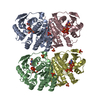
Entry Database : PDB / ID : 5yewTitle Structural basis for GTP hydrolysis and conformational change of mitofusin 1 in mediating mitochondrial fusion (Mitofusin-1,Mitofusin-1 fusion ...) x 2 Keywords / / / Function / homology Biological species Homo sapiens (human)Method / / / Resolution : 3.2 Å Authors Yan, L. / Qi, Y. / Huang, X. / Yu, C. Funding support Organization Grant number Country the National Natural Science Foundation of China 31700659 the National Natural Science Foundation of China 31225006 the National Natural Science Foundation of China 81322023
Journal : Nat. Struct. Mol. Biol. / Year : 2018Title : Structural basis for GTP hydrolysis and conformational change of MFN1 in mediating membrane fusionAuthors : Yan, L. / Qi, Y. / Huang, X. / Yu, C. / Lan, L. / Guo, X. / Rao, Z. / Hu, J. / Lou, Z. History Deposition Sep 20, 2017 Deposition site / Processing site Revision 1.0 Jan 31, 2018 Provider / Type Revision 1.1 Feb 28, 2018 Group / Category Item _citation.country / _citation.journal_abbrev ... _citation.country / _citation.journal_abbrev / _citation.journal_id_CSD / _citation.journal_id_ISSN / _citation.pdbx_database_id_DOI / _citation.year Revision 1.2 Mar 14, 2018 Group / Category Item / _citation.pdbx_database_id_PubMed / _citation.titleRevision 1.3 Mar 21, 2018 Group / Category Item / _citation.page_first / _citation.page_lastRevision 1.4 Mar 6, 2024 Group Data collection / Database references ... Data collection / Database references / Derived calculations / Refinement description Category chem_comp_atom / chem_comp_bond ... chem_comp_atom / chem_comp_bond / database_2 / pdbx_struct_conn_angle / refine_hist / software / struct_conn / struct_ncs_dom_lim Item _database_2.pdbx_DOI / _database_2.pdbx_database_accession ... _database_2.pdbx_DOI / _database_2.pdbx_database_accession / _pdbx_struct_conn_angle.ptnr1_auth_asym_id / _pdbx_struct_conn_angle.ptnr1_auth_comp_id / _pdbx_struct_conn_angle.ptnr1_auth_seq_id / _pdbx_struct_conn_angle.ptnr1_label_asym_id / _pdbx_struct_conn_angle.ptnr1_label_atom_id / _pdbx_struct_conn_angle.ptnr1_label_comp_id / _pdbx_struct_conn_angle.ptnr1_label_seq_id / _pdbx_struct_conn_angle.ptnr2_auth_asym_id / _pdbx_struct_conn_angle.ptnr2_auth_comp_id / _pdbx_struct_conn_angle.ptnr2_auth_seq_id / _pdbx_struct_conn_angle.ptnr2_label_asym_id / _pdbx_struct_conn_angle.ptnr2_label_atom_id / _pdbx_struct_conn_angle.ptnr2_label_comp_id / _pdbx_struct_conn_angle.ptnr3_auth_asym_id / _pdbx_struct_conn_angle.ptnr3_auth_comp_id / _pdbx_struct_conn_angle.ptnr3_auth_seq_id / _pdbx_struct_conn_angle.ptnr3_label_asym_id / _pdbx_struct_conn_angle.ptnr3_label_atom_id / _pdbx_struct_conn_angle.ptnr3_label_comp_id / _pdbx_struct_conn_angle.ptnr3_label_seq_id / _pdbx_struct_conn_angle.value / _refine_hist.d_res_low / _software.classification / _software.name / _struct_conn.pdbx_dist_value / _struct_conn.ptnr1_auth_asym_id / _struct_conn.ptnr1_auth_comp_id / _struct_conn.ptnr1_auth_seq_id / _struct_conn.ptnr1_label_asym_id / _struct_conn.ptnr1_label_atom_id / _struct_conn.ptnr1_label_comp_id / _struct_conn.ptnr1_label_seq_id / _struct_conn.ptnr2_auth_asym_id / _struct_conn.ptnr2_auth_comp_id / _struct_conn.ptnr2_auth_seq_id / _struct_conn.ptnr2_label_asym_id / _struct_conn.ptnr2_label_atom_id / _struct_conn.ptnr2_label_comp_id / _struct_ncs_dom_lim.beg_auth_comp_id / _struct_ncs_dom_lim.beg_label_asym_id / _struct_ncs_dom_lim.beg_label_comp_id / _struct_ncs_dom_lim.beg_label_seq_id / _struct_ncs_dom_lim.end_auth_comp_id / _struct_ncs_dom_lim.end_label_asym_id / _struct_ncs_dom_lim.end_label_comp_id / _struct_ncs_dom_lim.end_label_seq_id
Show all Show less
 Movie
Movie Controller

 Yorodumi
 Open data
 Basic information
 Components
 Keywords
 HYDROLASE /
  Function and homology information
 Authors
 China, 3items
  Citation
 Structure visualization
 Downloads & links
 Other downloads


 PDBj
PDBj


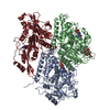
 Assembly
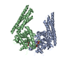


 Type: RNA linking / Mass: 443.201 Da / Num. of mol.: 3 / Source method: obtained synthetically / Formula: C10H15N5O11P2 / Comment: GDP, energy-carrying molecule*YM
 Mass: 66.007 Da / Num. of mol.: 3 / Source method: obtained synthetically / Formula: BeF3
 Mass: 39.098 Da / Num. of mol.: 3 / Source method: obtained synthetically / Formula: K
 Mass: 24.305 Da / Num. of mol.: 3 / Source method: obtained synthetically / Formula: Mg
 Sample preparation
 Processing