 Movie
Movie Controller

[English] 日本語
 Yorodumi
- PDB-4c5m: Structure of the pyridoxal kinase from Staphylococcus aureus in c... -
+ Open data

- Basic information
| Entry | Database: PDB / ID: 4c5m | ||||||
|---|---|---|---|---|---|---|---|
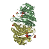
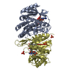
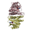
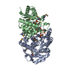
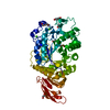
| Title | Structure of the pyridoxal kinase from Staphylococcus aureus in complex with AMP-PCP | ||||||
 Components Components | PHOSPHOMETHYLPYRIMIDINE KINASE | ||||||
 Keywords Keywords | TRANSFERASE / RIBOKINASE | ||||||
| Function / homology |  Function and homology informationphosphomethylpyrimidine kinase activity / pyridoxal kinase / thiamine biosynthetic process / phosphorylation / nucleotide binding Function and homology informationphosphomethylpyrimidine kinase activity / pyridoxal kinase / thiamine biosynthetic process / phosphorylation / nucleotide bindingSimilarity search - Function | ||||||
| Biological species |  STAPHYLOCOCCUS AUREUS SUBSP. AUREUS MU50 (bacteria) STAPHYLOCOCCUS AUREUS SUBSP. AUREUS MU50 (bacteria) | ||||||
| Method | X-RAY DIFFRACTION / SYNCHROTRON / MOLECULAR REPLACEMENT / Resolution: 1.45 Å | ||||||
 Authors Authors | Nodwell, M. / Alte, F. / Sieber, S.A. / Schneider, S. | ||||||
 Citation Citation | Journal: J.Am.Chem.Soc. / Year: 2014 Title: A Subfamily of Bacterial Ribokinases Utilizes a Hemithioacetal for Pyridoxal Phosphate Salvage. Authors: Nodwell, M.B. / Koch, M.F. / Alte, F. / Schneider, S. / Sieber, S.A. | ||||||
| History |
|
- Structure visualization
| Structure viewer | Molecule: MolmilJmol/JSmol |
|---|
- Downloads & links
-Download
| PDBx/mmCIF format | 4c5m.cif.gz | 480.8 KB | Display | PDBx/mmCIF format |
|---|---|---|---|---|
| PDB format | pdb4c5m.ent.gz | 397.6 KB | Display | PDB format |
| PDBx/mmJSON format | 4c5m.json.gz | Tree view | PDBx/mmJSON format | |
| Others |  Other downloads Other downloads |
-Validation report
| Arichive directory | https://data.pdbj.org/pub/pdb/validation_reports/c5/4c5mftp://data.pdbj.org/pub/pdb/validation_reports/c5/4c5m | HTTPS FTP |
|---|
-Related structure data
| Related structure data |  4c5jSC  4c5kC  4c5lC  4c5nC S: Starting model for refinement C: citing same article ( |
|---|---|
| Similar structure data |


-Links
 PDBj
PDBj


- Assembly
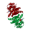
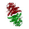
| Deposited unit | 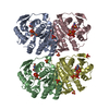
| ||||||||||||||||
|---|---|---|---|---|---|---|---|---|---|---|---|---|---|---|---|---|---|
| 1 | 
| ||||||||||||||||
| 2 | 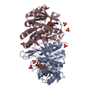
| ||||||||||||||||
| Unit cell |
| ||||||||||||||||
| Noncrystallographic symmetry (NCS) | NCS oper:
|
-Components
| #1: Protein | / PYRIDOXAL KINASE Mass: 29811.793 Da / Num. of mol.: 4 Source method: isolated from a genetically manipulated source Source: (gene. exp.) STAPHYLOCOCCUS AUREUS SUBSP. AUREUS MU50 (bacteria)Production host: ESCHERICHIA COLI (E. coli)References: UniProt: Q99W31, UniProt: A0A0H3JTP0*PLUS, pyridoxal kinase#2: Chemical | ChemComp-ACP /   Mass: 505.208 Da / Num. of mol.: 4 / Source method: obtained synthetically / Formula: C11H18N5O12P3 / Comment: AMP-PCP, energy-carrying molecule analogue*YM Mass: 505.208 Da / Num. of mol.: 4 / Source method: obtained synthetically / Formula: C11H18N5O12P3 / Comment: AMP-PCP, energy-carrying molecule analogue*YM#3: Chemical | ChemComp-SO4 / Sulfate  Mass: 96.063 Da / Num. of mol.: 17 / Source method: obtained synthetically / Formula: SO4 Mass: 96.063 Da / Num. of mol.: 17 / Source method: obtained synthetically / Formula: SO4#4: Water | ChemComp-HOH / | Water Mass: 18.015 Da / Num. of mol.: 978 / Source method: isolated from a natural source / Formula: H2O Mass: 18.015 Da / Num. of mol.: 978 / Source method: isolated from a natural source / Formula: H2OSequence details | FIRST AMINOACID IS A GLY INSTEAD OF MET DUE TO REMOVAL OF THE AFFINITY TAG | |
|---|
-Experimental details
-Experiment
| Experiment | Method: X-RAY DIFFRACTION / Number of used crystals: 1 |
|---|
- Sample preparation
| Crystal | Density Matthews: 2.19 Å3/Da / Density % sol: 45 % / Description: NONE |
|---|---|
| Crystal grow | Details: 50MM HEPES, 2M NH4SO4 |
-Data collection
| Diffraction | Mean temperature: 100 K |
|---|---|
| Diffraction source | Source: SYNCHROTRON / Site: SLS  / Beamline: X06DA / Wavelength: 1 / Beamline: X06DA / Wavelength: 1 |
| Detector | Type: DECTRIS PILATUS / Detector: PIXEL / Date: Jul 18, 2013 |
| Radiation | Protocol: SINGLE WAVELENGTH / Monochromatic (M) / Laue (L): M / Scattering type: x-ray |
| Radiation wavelength | Wavelength: 1 Å / Relative weight: 1 |
| Reflection | Resolution: 1.45→48.76 Å / Num. obs: 185724 / % possible obs: 99.5 % / Observed criterion σ(I): 1.5 / Redundancy: 8.8 % / Rmerge(I) obs: 0.06 / Net I/σ(I): 20.38 |
| Reflection shell | Resolution: 1.45→1.5 Å / Redundancy: 8.5 % / Rmerge(I) obs: 1.17 / Mean I/σ(I) obs: 1.79 / % possible all: 96.3 |
- Processing
| Software |
| ||||||||||||||||||||||||||||||||||||||||||||||||||||||||||||||||||||||||||||||||||||||||||||||||||||||||||||||||||||||||||||||||||||||||||||||||||||||||||||||||||||||||||||||||||||||
|---|---|---|---|---|---|---|---|---|---|---|---|---|---|---|---|---|---|---|---|---|---|---|---|---|---|---|---|---|---|---|---|---|---|---|---|---|---|---|---|---|---|---|---|---|---|---|---|---|---|---|---|---|---|---|---|---|---|---|---|---|---|---|---|---|---|---|---|---|---|---|---|---|---|---|---|---|---|---|---|---|---|---|---|---|---|---|---|---|---|---|---|---|---|---|---|---|---|---|---|---|---|---|---|---|---|---|---|---|---|---|---|---|---|---|---|---|---|---|---|---|---|---|---|---|---|---|---|---|---|---|---|---|---|---|---|---|---|---|---|---|---|---|---|---|---|---|---|---|---|---|---|---|---|---|---|---|---|---|---|---|---|---|---|---|---|---|---|---|---|---|---|---|---|---|---|---|---|---|---|---|---|---|---|
| Refinement | Method to determine structure: MOLECULAR REPLACEMENT Starting model: PDB ENTRY 4C5J Resolution: 1.45→48.76 Å / Cor.coef. Fo:Fc: 0.982 / Cor.coef. Fo:Fc free: 0.972 / SU B: 2.323 / SU ML: 0.039 / Cross valid method: THROUGHOUT / ESU R: 0.06 / ESU R Free: 0.059 / Stereochemistry target values: MAXIMUM LIKELIHOOD Details: HYDROGENS HAVE BEEN ADDED IN THE RIDING POSITIONS. U VALUES REFINED INDIVIDUALLY
| ||||||||||||||||||||||||||||||||||||||||||||||||||||||||||||||||||||||||||||||||||||||||||||||||||||||||||||||||||||||||||||||||||||||||||||||||||||||||||||||||||||||||||||||||||||||
| Solvent computation | Ion probe radii: 0.8 Å / Shrinkage radii: 0.8 Å / VDW probe radii: 1.2 Å / Solvent model: MASK | ||||||||||||||||||||||||||||||||||||||||||||||||||||||||||||||||||||||||||||||||||||||||||||||||||||||||||||||||||||||||||||||||||||||||||||||||||||||||||||||||||||||||||||||||||||||
| Displacement parameters | Biso mean: 21.882 Å2
| ||||||||||||||||||||||||||||||||||||||||||||||||||||||||||||||||||||||||||||||||||||||||||||||||||||||||||||||||||||||||||||||||||||||||||||||||||||||||||||||||||||||||||||||||||||||
| Refinement step | Cycle: LAST / Resolution: 1.45→48.76 Å
| ||||||||||||||||||||||||||||||||||||||||||||||||||||||||||||||||||||||||||||||||||||||||||||||||||||||||||||||||||||||||||||||||||||||||||||||||||||||||||||||||||||||||||||||||||||||
| Refine LS restraints |
|