 Movie
Movie Controller

[English] 日本語
 Yorodumi
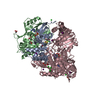
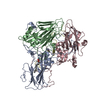
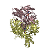
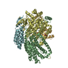
- PDB-5y6q: Crystal structure of an aldehyde oxidase from Methylobacillus sp.... -
+ Open data

- Basic information
| Entry | Database: PDB / ID: 5y6q | ||||||
|---|---|---|---|---|---|---|---|
| Title | Crystal structure of an aldehyde oxidase from Methylobacillus sp. KY4400 | ||||||
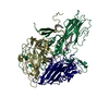
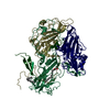
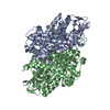
 Components Components | (Aldehyde oxidase ... ) x 3 ) x 3 | ||||||
 Keywords Keywords | OXIDOREDUCTASE / aldehyde oxidase molybdenum enzyme Methylobacillus sp. KY4400 | ||||||
| Function / homology |  Function and homology information Function and homology informationFAD binding / 2 iron, 2 sulfur cluster binding / electron transfer activity / oxidoreductase activity / metal ion bindingSimilarity search - Function | ||||||
| Biological species |  Methylobacillus sp. KY4400 (bacteria) Methylobacillus sp. KY4400 (bacteria) | ||||||
| Method | X-RAY DIFFRACTION / SYNCHROTRON / MOLECULAR REPLACEMENT / Resolution: 2.5 Å | ||||||
 Authors Authors | Mikami, B. / Uchida, H. | ||||||
| Funding support |  Japan, 1items Japan, 1items
| ||||||
 Citation Citation | Journal: J. Biochem. / Year: 2018 Title: Crystal structure of an aldehyde oxidase from Methylobacillus sp. KY4400. Authors: Uchida, H. / Mikami, B. / Yamane-Tanabe, A. / Ito, A. / Hirano, K. / Oki, M. #1: Journal: Biosci. Biotechnol. Biochem. / Year: 2005 Title: Cloning and sequencing of the aldehyde oxidase gene from Methylobacillus sp. KY4400. Authors: Yasuhara, A. / Akiba-Goto, M. / Aisaka, K. | ||||||
| History |
|
- Structure visualization
| Structure viewer | Molecule: MolmilJmol/JSmol |
|---|
- Downloads & links
-Download
| PDBx/mmCIF format | 5y6q.cif.gz | 269.1 KB | Display | PDBx/mmCIF format |
|---|---|---|---|---|
| PDB format | pdb5y6q.ent.gz | 207.3 KB | Display | PDB format |
| PDBx/mmJSON format | 5y6q.json.gz | Tree view | PDBx/mmJSON format | |
| Others |  Other downloads Other downloads |
-Validation report
| Arichive directory | https://data.pdbj.org/pub/pdb/validation_reports/y6/5y6qftp://data.pdbj.org/pub/pdb/validation_reports/y6/5y6q | HTTPS FTP |
|---|
-Related structure data
| Related structure data | 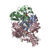 5g5hS S: Starting model for refinement |
|---|---|
| Similar structure data |


-Links
 PDBj
PDBj


- Assembly
| Deposited unit | 
| ||||||||
|---|---|---|---|---|---|---|---|---|---|
| 1 |
| ||||||||
| Unit cell |
|
-Components
-Aldehyde oxidase ... , 3 types, 3 molecules ABC
| #1: Protein | Mass: 17311.635 Da / Num. of mol.: 1 Source method: isolated from a genetically manipulated source Source: (gene. exp.) Methylobacillus sp. KY4400 (bacteria) / Gene: aoms / Production host: Escherichia coli K-12 (bacteria) / Strain (production host): K-12 / References: UniProt: Q84IY0 |
|---|---|
| #2: Protein | Mass: 35616.105 Da / Num. of mol.: 1 Source method: isolated from a genetically manipulated source Source: (gene. exp.) Methylobacillus sp. KY4400 (bacteria) / Gene: aomm / Production host: Escherichia coli K-12 (bacteria) / Strain (production host): K-12 / References: UniProt: Q84IX9 |
| #3: Protein | Mass: 83153.828 Da / Num. of mol.: 1 Source method: isolated from a genetically manipulated source Source: (gene. exp.) Methylobacillus sp. KY4400 (bacteria) / Gene: aoml / Production host: Escherichia coli K-12 (bacteria) / Strain (production host): K-12 / References: UniProt: Q84IX8 |
-Non-polymers , 9 types, 533 molecules 


| #4: Chemical | Iron–sulfur cluster Mass: 175.820 Da / Num. of mol.: 2 / Source method: obtained synthetically / Formula: Fe2S2 Mass: 175.820 Da / Num. of mol.: 2 / Source method: obtained synthetically / Formula: Fe2S2#5: Chemical | ChemComp-SO4 / Sulfate Mass: 96.063 Da / Num. of mol.: 12 / Source method: obtained synthetically / Formula: SO4 Mass: 96.063 Da / Num. of mol.: 12 / Source method: obtained synthetically / Formula: SO4#6: Chemical | ChemComp-FAD / | Flavin adenine dinucleotide Mass: 785.550 Da / Num. of mol.: 1 / Source method: obtained synthetically / Formula: C27H33N9O15P2 / Comment: FAD*YM Mass: 785.550 Da / Num. of mol.: 1 / Source method: obtained synthetically / Formula: C27H33N9O15P2 / Comment: FAD*YM#7: Chemical | ChemComp-SF4 / | Iron–sulfur cluster Mass: 351.640 Da / Num. of mol.: 1 / Source method: obtained synthetically / Formula: Fe4S4 Mass: 351.640 Da / Num. of mol.: 1 / Source method: obtained synthetically / Formula: Fe4S4#8: Chemical | ChemComp-GOL / Glycerol Mass: 92.094 Da / Num. of mol.: 6 / Source method: obtained synthetically / Formula: C3H8O3 Mass: 92.094 Da / Num. of mol.: 6 / Source method: obtained synthetically / Formula: C3H8O3#9: Chemical | ChemComp-MOS / |  Mass: 161.012 Da / Num. of mol.: 1 / Source method: obtained synthetically / Formula: HMoO2S Mass: 161.012 Da / Num. of mol.: 1 / Source method: obtained synthetically / Formula: HMoO2S#10: Chemical | ChemComp-MCN / |  Mass: 696.501 Da / Num. of mol.: 1 / Source method: obtained synthetically / Formula: C19H22N8O13P2S2 Mass: 696.501 Da / Num. of mol.: 1 / Source method: obtained synthetically / Formula: C19H22N8O13P2S2#11: Chemical | Isopropyl alcohol Mass: 60.095 Da / Num. of mol.: 2 / Source method: obtained synthetically / Formula: C3H8O / Comment: alkaloid*YM Mass: 60.095 Da / Num. of mol.: 2 / Source method: obtained synthetically / Formula: C3H8O / Comment: alkaloid*YM#12: Water | ChemComp-HOH / | WaterMass: 18.015 Da / Num. of mol.: 507 / Source method: isolated from a natural source / Formula: H2O |
|---|
-Details
| Sequence details | The depositors state that the database UNP codes Q84IY0/Q84IX9 are incorrect at these positions. |
|---|
-Experimental details
-Experiment
| Experiment | Method: X-RAY DIFFRACTION / Number of used crystals: 1 |
|---|
- Sample preparation
| Crystal | Density Matthews: 2.3 Å3/Da / Density % sol: 46 % |
|---|---|
| Crystal grow | Temperature: 293 K / Method: vapor diffusion, hanging drop / pH: 5.5 Details: 0.1 M Sodium citrate buffer, pH 5.5, 4-5% W/V isopropanol, 22-27% W/V PEG 4000, 10-16% W/V ammonium sulfate, red cubic flash cooled after brief soaking to the bottom solution containing 30% glycerol |
-Data collection
| Diffraction | Mean temperature: 100 K |
|---|---|
| Diffraction source | Source: SYNCHROTRON / Site: SPring-8 / Beamline: BL38B1 / Wavelength: 1 Å |
| Detector | Type: RIGAKU JUPITER 210 / Detector: CCD / Date: Oct 8, 2004 |
| Radiation | Protocol: SINGLE WAVELENGTH / Monochromatic (M) / Laue (L): M / Scattering type: x-ray |
| Radiation wavelength | Wavelength: 1 Å / Relative weight: 1 |
| Reflection | Resolution: 2.5→50 Å / Num. obs: 44006 / % possible obs: 99.1 % / Redundancy: 15.1 % / Biso Wilson estimate: 26.8 Å2 / Rmerge(I) obs: 0.087 / Rpim(I) all: 0.022 / Χ2: 0.856 / Net I/σ(I): 24.5 |
| Reflection shell | Resolution: 2.5→2.54 Å / Redundancy: 5.7 % / Rmerge(I) obs: 0.361 / Mean I/σ(I) obs: 3.13 / Num. unique obs: 1944 / CC1/2: 0.901 / Rpim(I) all: 0.161 / Χ2: 0.487 / % possible all: 90.7 |
- Processing
| Software |
| |||||||||||||||||||||||||||||||||||||||||||||||||||||||||||||||||||||||||||||||||||||||||||||||||||||||||||||||||||||||
|---|---|---|---|---|---|---|---|---|---|---|---|---|---|---|---|---|---|---|---|---|---|---|---|---|---|---|---|---|---|---|---|---|---|---|---|---|---|---|---|---|---|---|---|---|---|---|---|---|---|---|---|---|---|---|---|---|---|---|---|---|---|---|---|---|---|---|---|---|---|---|---|---|---|---|---|---|---|---|---|---|---|---|---|---|---|---|---|---|---|---|---|---|---|---|---|---|---|---|---|---|---|---|---|---|---|---|---|---|---|---|---|---|---|---|---|---|---|---|---|---|
| Refinement | Method to determine structure: MOLECULAR REPLACEMENT Starting model: 5G5H Resolution: 2.5→46.116 Å / SU ML: 0.27 / Cross valid method: FREE R-VALUE / σ(F): 1.35 / Phase error: 21.79 / Stereochemistry target values: ML
| |||||||||||||||||||||||||||||||||||||||||||||||||||||||||||||||||||||||||||||||||||||||||||||||||||||||||||||||||||||||
| Solvent computation | Shrinkage radii: 0.9 Å / VDW probe radii: 1.11 Å / Solvent model: FLAT BULK SOLVENT MODEL | |||||||||||||||||||||||||||||||||||||||||||||||||||||||||||||||||||||||||||||||||||||||||||||||||||||||||||||||||||||||
| Refinement step | Cycle: LAST / Resolution: 2.5→46.116 Å
| |||||||||||||||||||||||||||||||||||||||||||||||||||||||||||||||||||||||||||||||||||||||||||||||||||||||||||||||||||||||
| Refine LS restraints |
| |||||||||||||||||||||||||||||||||||||||||||||||||||||||||||||||||||||||||||||||||||||||||||||||||||||||||||||||||||||||
| LS refinement shell |
|
