 Movie
Movie Controller


+ Open data

- Basic information
| Entry | Database: PDB / ID: 5y2d | ||||||
|---|---|---|---|---|---|---|---|
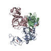
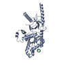
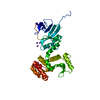
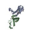
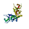
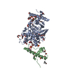
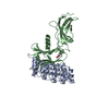
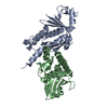
| Title | Crystal structure of H. pylori HtrA | ||||||
 Components Components |
| ||||||
 Keywords Keywords |  HYDROLASE / Serine protease HYDROLASE / Serine protease | ||||||
| Function / homology |  Function and homology informationpeptidase Do / metalloendopeptidase activity / periplasmic space / serine-type endopeptidase activity / proteolysis Function and homology informationpeptidase Do / metalloendopeptidase activity / periplasmic space / serine-type endopeptidase activity / proteolysisSimilarity search - Function | ||||||
| Biological species |   Helicobacter pylori (bacteria)Helicobacter pylori 26695 (bacteria) Helicobacter pylori (bacteria)Helicobacter pylori 26695 (bacteria) | ||||||
| Method | X-RAY DIFFRACTION / SYNCHROTRON / Resolution: 3.70009854828 Å | ||||||
 Authors Authors | Zhang, Z. / Huang, Q. / Tao, X. | ||||||
 Citation Citation | Journal: J.Biol.Chem. / Year: 2019 Title: The unique trimeric assembly of the virulence factor HtrA fromHelicobacter pylorioccurs via N-terminal domain swapping. Authors: Zhang, Z. / Huang, Q. / Tao, X. / Song, G. / Zheng, P. / Li, H. / Sun, H. / Xia, W. | ||||||
| History |
|
- Structure visualization
| Structure viewer | Molecule: MolmilJmol/JSmol |
|---|
- Downloads & links
-Download
| PDBx/mmCIF format | 5y2d.cif.gz | 68.4 KB | Display | PDBx/mmCIF format |
|---|---|---|---|---|
| PDB format | pdb5y2d.ent.gz | 45.6 KB | Display | PDB format |
| PDBx/mmJSON format | 5y2d.json.gz | Tree view | PDBx/mmJSON format | |
| Others |  Other downloads Other downloads |
-Validation report
| Arichive directory | https://data.pdbj.org/pub/pdb/validation_reports/y2/5y2dftp://data.pdbj.org/pub/pdb/validation_reports/y2/5y2d | HTTPS FTP |
|---|
-Related structure data


-Links
 PDBj
PDBj

- Assembly
| Deposited unit | 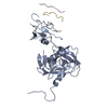
| ||||||||||
|---|---|---|---|---|---|---|---|---|---|---|---|
| 1 |
| ||||||||||
| Unit cell |
|
-Components
-Protein , 1 types, 1 molecules A
| #1: Protein | Mass: 50370.898 Da / Num. of mol.: 1 Source method: isolated from a genetically manipulated source Source: (gene. exp.) Helicobacter pylori (bacteria) / Strain: ATCC 700392 / 26695 / Gene: hp1018/19 / Production host: Escherichia coli BL21 (bacteria) / Strain (production host): BL21 / References: UniProt: G2J5T2, peptidase Do |
|---|
-Protein/peptide , 3 types, 3 molecules BCD
| #2: Protein/peptide | Mass: 231.249 Da / Num. of mol.: 1 Source method: isolated from a genetically manipulated source Source: (gene. exp.) Helicobacter pylori 26695 (bacteria) / Strain: 26695 / Production host: Escherichia coli (E. coli) |
|---|---|
| #3: Protein/peptide | Mass: 373.404 Da / Num. of mol.: 1 Source method: isolated from a genetically manipulated source Source: (gene. exp.) Helicobacter pylori 26695 (bacteria) / Strain: 26695 / Production host: Escherichia coli (E. coli) |
| #4: Protein/peptide | Mass: 786.895 Da / Num. of mol.: 1 Source method: isolated from a genetically manipulated source Source: (gene. exp.) Helicobacter pylori 26695 (bacteria) / Strain: 26695 / Production host: Escherichia coli (E. coli) |
-UNK-UNK-UNK-UNK-UNK-UNK-UNK- ... , 2 types, 2 molecules EF
| #5: Protein/peptide | Mass: 586.638 Da / Num. of mol.: 1 Source method: isolated from a genetically manipulated source Source: (gene. exp.) Helicobacter pylori 26695 (bacteria) / Strain: 26695 / Production host: Escherichia coli (E. coli) |
|---|---|
| #6: Protein/peptide | Mass: 799.871 Da / Num. of mol.: 1 Source method: isolated from a genetically manipulated source Source: (gene. exp.) Helicobacter pylori 26695 (bacteria) / Strain: 26695 / Production host: Escherichia coli (E. coli) |
-Experimental details
-Experiment
| Experiment | Method: X-RAY DIFFRACTION / Number of used crystals: 1 |
|---|
- Sample preparation
| Crystal | Density Matthews: 2.72 Å3/Da / Density % sol: 54.75 % |
|---|---|
| Crystal grow | Temperature: 293 K / Method: vapor diffusion, hanging drop / pH: 7 / Details: 2.1 M DL-malic acid pH 7.0, 0.1 M HEPES pH 7.0 |
-Data collection
| Diffraction | Mean temperature: 100 K | |||||||||||||||||||||||||||||||||||||||||||||||||||||||||||||||||||||||||||||||||||||||||||||||||||||||||||||||||||||||||||||||||||||||
|---|---|---|---|---|---|---|---|---|---|---|---|---|---|---|---|---|---|---|---|---|---|---|---|---|---|---|---|---|---|---|---|---|---|---|---|---|---|---|---|---|---|---|---|---|---|---|---|---|---|---|---|---|---|---|---|---|---|---|---|---|---|---|---|---|---|---|---|---|---|---|---|---|---|---|---|---|---|---|---|---|---|---|---|---|---|---|---|---|---|---|---|---|---|---|---|---|---|---|---|---|---|---|---|---|---|---|---|---|---|---|---|---|---|---|---|---|---|---|---|---|---|---|---|---|---|---|---|---|---|---|---|---|---|---|---|---|
| Diffraction source | Source: SYNCHROTRON / Site: SSRF  / Beamline: BL17B1 / Wavelength: 0.99 Å / Beamline: BL17B1 / Wavelength: 0.99 Å | |||||||||||||||||||||||||||||||||||||||||||||||||||||||||||||||||||||||||||||||||||||||||||||||||||||||||||||||||||||||||||||||||||||||
| Detector | Type: ADSC QUANTUM 315r / Detector: CCD / Date: Oct 27, 2016 | |||||||||||||||||||||||||||||||||||||||||||||||||||||||||||||||||||||||||||||||||||||||||||||||||||||||||||||||||||||||||||||||||||||||
| Radiation | Protocol: SINGLE WAVELENGTH / Monochromatic (M) / Laue (L): M / Scattering type: x-ray | |||||||||||||||||||||||||||||||||||||||||||||||||||||||||||||||||||||||||||||||||||||||||||||||||||||||||||||||||||||||||||||||||||||||
| Radiation wavelength | Wavelength: 0.99 Å / Relative weight: 1 | |||||||||||||||||||||||||||||||||||||||||||||||||||||||||||||||||||||||||||||||||||||||||||||||||||||||||||||||||||||||||||||||||||||||
| Reflection | Resolution: 3.7→50 Å / Num. obs: 6505 / % possible obs: 100 % / Redundancy: 11.1 % / Biso Wilson estimate: 63.8079972517 Å2 / Rmerge(I) obs: 0.117 / Rpim(I) all: 0.037 / Rrim(I) all: 0.123 / Χ2: 1.04 / Net I/σ(I): 5 | |||||||||||||||||||||||||||||||||||||||||||||||||||||||||||||||||||||||||||||||||||||||||||||||||||||||||||||||||||||||||||||||||||||||
| Reflection shell | Diffraction-ID: 1
|
- Processing
| Software |
| |||||||||||||||||||||||||||||||||||
|---|---|---|---|---|---|---|---|---|---|---|---|---|---|---|---|---|---|---|---|---|---|---|---|---|---|---|---|---|---|---|---|---|---|---|---|---|
| Refinement | Resolution: 3.70009854828→44.4946700101 Å / SU ML: 0.468111806412 / Cross valid method: FREE R-VALUE / σ(F): 1.35739823429 / Phase error: 34.0435691982 Stereochemistry target values: GeoStd + Monomer Library + CDL v1.2
| |||||||||||||||||||||||||||||||||||
| Solvent computation | Shrinkage radii: 0.9 Å / VDW probe radii: 1.11 Å / Solvent model: FLAT BULK SOLVENT MODEL | |||||||||||||||||||||||||||||||||||
| Displacement parameters | Biso mean: 49.0221781202 Å2 | |||||||||||||||||||||||||||||||||||
| Refinement step | Cycle: LAST / Resolution: 3.70009854828→44.4946700101 Å
| |||||||||||||||||||||||||||||||||||
| Refine LS restraints |
| |||||||||||||||||||||||||||||||||||
| LS refinement shell |
|
