 Movie
Movie Controller


+ Open data

- Basic information
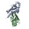
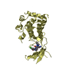
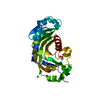
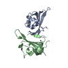
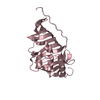
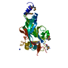
| Entry | Database: PDB / ID: 3vnf | ||||||
|---|---|---|---|---|---|---|---|
| Title | Structure of the ebolavirus protein VP24 from Sudan | ||||||
 Components Components | Membrane-associated protein VP24 | ||||||
 Keywords Keywords |  VIRAL PROTEIN / ebolavirus / interferon antagonist / VP24 / STAT1 / Zaire / Sudan / Reston / VP35 / karyopherin alpha / IFN response pathway VIRAL PROTEIN / ebolavirus / interferon antagonist / VP24 / STAT1 / Zaire / Sudan / Reston / VP35 / karyopherin alpha / IFN response pathway | ||||||
| Function / homology |  Function and homology information Function and homology informationhost cell endomembrane system / endomembrane system / virus-mediated perturbation of host defense response / host cell plasma membrane / virion membrane / structural molecule activity / membrane / plasma membraneSimilarity search - Function | ||||||
| Biological species |  Sudan ebolavirus Sudan ebolavirus | ||||||
| Method | X-RAY DIFFRACTION / SYNCHROTRON / MAD / Resolution: 2.1 Å | ||||||
 Authors Authors | Zhang, A.P.P. | ||||||
 Citation Citation | Journal: Plos Pathog. / Year: 2012 Title: The ebola virus interferon antagonist VP24 directly binds STAT1 and has a novel, pyramidal fold Authors: Zhang, A.P.P. / Bornholdt, Z.A. / Liu, T. / Abelson, D.M. / Lee, D.E. / Li, S. / Woods Jr., V.L. / Saphire, E.O. | ||||||
| History |
|
- Structure visualization
| Structure viewer | Molecule: MolmilJmol/JSmol |
|---|
- Downloads & links
-Download
| PDBx/mmCIF format | 3vnf.cif.gz | 95.3 KB | Display | PDBx/mmCIF format |
|---|---|---|---|---|
| PDB format | pdb3vnf.ent.gz | 73.4 KB | Display | PDB format |
| PDBx/mmJSON format | 3vnf.json.gz | Tree view | PDBx/mmJSON format | |
| Others |  Other downloads Other downloads |
-Validation report
| Arichive directory | https://data.pdbj.org/pub/pdb/validation_reports/vn/3vnfftp://data.pdbj.org/pub/pdb/validation_reports/vn/3vnf | HTTPS FTP |
|---|
-Related structure data


-Links
 PDBj
PDBj
- Assembly
| Deposited unit | 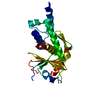
| ||||||||
|---|---|---|---|---|---|---|---|---|---|
| 1 |
| ||||||||
| 2 | 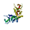
| ||||||||
| Unit cell |
|
-Components
| #1: Protein | Mass: 24583.521 Da / Num. of mol.: 1 / Fragment: UNP residues 13-228 / Mutation: V22A Source method: isolated from a genetically manipulated source Source: (gene. exp.) Sudan ebolavirus / Gene: VP24 / Production host:  Escherichia coli (E. coli) / References: UniProt: B0LPM0, UniProt: Q5XX02*PLUS Escherichia coli (E. coli) / References: UniProt: B0LPM0, UniProt: Q5XX02*PLUS |
|---|---|
| #2: Water | ChemComp-HOH / Water Mass: 18.015 Da / Num. of mol.: 46 / Source method: isolated from a natural source / Formula: H2O Mass: 18.015 Da / Num. of mol.: 46 / Source method: isolated from a natural source / Formula: H2O |
-Experimental details
-Experiment
| Experiment | Method: X-RAY DIFFRACTION / Number of used crystals: 2 |
|---|
- Sample preparation
| Crystal |
| |||||||||||||||
|---|---|---|---|---|---|---|---|---|---|---|---|---|---|---|---|---|
| Crystal grow |
|
-Data collection
| Diffraction |
| ||||||||||||||||||
|---|---|---|---|---|---|---|---|---|---|---|---|---|---|---|---|---|---|---|---|
| Diffraction source |
| ||||||||||||||||||
| Detector |
| ||||||||||||||||||
| Radiation |
| ||||||||||||||||||
| Radiation wavelength |
| ||||||||||||||||||
| Reflection | Resolution: 1.8→33.03 Å / Num. all: 25038 / Num. obs: 23986 / % possible obs: 95.8 % / Observed criterion σ(F): 0 / Observed criterion σ(I): 0 / Redundancy: 8.3 % / Biso Wilson estimate: 53.67 Å2 / Rmerge(I) obs: 0.075 / Net I/σ(I): 8.3 | ||||||||||||||||||
| Reflection shell | Highest resolution: 1.8 Å / % possible all: 95.8 |

-Phasing
| Phasing | Method: MAD |
|---|
- Processing
| Software |
| |||||||||||||||||||||||||||||||||||||||||||||||||||||||||||||||||||||||||||
|---|---|---|---|---|---|---|---|---|---|---|---|---|---|---|---|---|---|---|---|---|---|---|---|---|---|---|---|---|---|---|---|---|---|---|---|---|---|---|---|---|---|---|---|---|---|---|---|---|---|---|---|---|---|---|---|---|---|---|---|---|---|---|---|---|---|---|---|---|---|---|---|---|---|---|---|---|
| Refinement | Method to determine structure: MAD / Resolution: 2.1→33.03 Å / Occupancy max: 1 / Occupancy min: 1 / FOM work R set: 0.7717 / SU ML: 0.38 / σ(F): 0 / Phase error: 28.93 / Stereochemistry target values: ML
| |||||||||||||||||||||||||||||||||||||||||||||||||||||||||||||||||||||||||||
| Solvent computation | Shrinkage radii: 0.83 Å / VDW probe radii: 1.1 Å / Solvent model: FLAT BULK SOLVENT MODEL / Bsol: 70.101 Å2 / ksol: 0.333 e/Å3 | |||||||||||||||||||||||||||||||||||||||||||||||||||||||||||||||||||||||||||
| Displacement parameters | Biso max: 193.9 Å2 / Biso mean: 77.1694 Å2 / Biso min: 30.9 Å2
| |||||||||||||||||||||||||||||||||||||||||||||||||||||||||||||||||||||||||||
| Refinement step | Cycle: LAST / Resolution: 2.1→33.03 Å
| |||||||||||||||||||||||||||||||||||||||||||||||||||||||||||||||||||||||||||
| Refine LS restraints |
| |||||||||||||||||||||||||||||||||||||||||||||||||||||||||||||||||||||||||||
| LS refinement shell | Refine-ID: X-RAY DIFFRACTION / Total num. of bins used: 9
| |||||||||||||||||||||||||||||||||||||||||||||||||||||||||||||||||||||||||||
| Refinement TLS params. | Method: refined / Refine-ID: X-RAY DIFFRACTION
| |||||||||||||||||||||||||||||||||||||||||||||||||||||||||||||||||||||||||||
| Refinement TLS group |
|
