 Movie
Movie Controller

[English] 日本語
 Yorodumi
- PDB-5xz9: Crystal Structure of Phosphofructokinase from Staphylococcus aure... -
+ Open data

- Basic information
| Entry | Database: PDB / ID: 5xz9 | ||||||
|---|---|---|---|---|---|---|---|
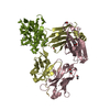
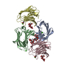
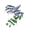
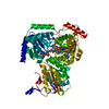
| Title | Crystal Structure of Phosphofructokinase from Staphylococcus aureus in complex with adenylylimidodiphosphate, the ATP analogue | ||||||
 Components Components | ATP-dependent 6-phosphofructokinase | ||||||
 Keywords Keywords |  TRANSFERASE / Phosphofructokinase / Staphylococcus aureus TRANSFERASE / Phosphofructokinase / Staphylococcus aureus | ||||||
| Function / homology |  Function and homology information6-phosphofructokinase complex / 6-phosphofructokinase / 6-phosphofructokinase activity / fructose-6-phosphate binding / fructose 6-phosphate metabolic process / canonical glycolysis / fructose 1,6-bisphosphate metabolic process / monosaccharide binding / AMP binding / ATP binding ...6-phosphofructokinase complex / 6-phosphofructokinase / 6-phosphofructokinase activity / fructose-6-phosphate binding / fructose 6-phosphate metabolic process / canonical glycolysis / fructose 1,6-bisphosphate metabolic process / monosaccharide binding / AMP binding / ATP binding / membrane / identical protein binding / metal ion binding Function and homology information6-phosphofructokinase complex / 6-phosphofructokinase / 6-phosphofructokinase activity / fructose-6-phosphate binding / fructose 6-phosphate metabolic process / canonical glycolysis / fructose 1,6-bisphosphate metabolic process / monosaccharide binding / AMP binding / ATP binding ...6-phosphofructokinase complex / 6-phosphofructokinase / 6-phosphofructokinase activity / fructose-6-phosphate binding / fructose 6-phosphate metabolic process / canonical glycolysis / fructose 1,6-bisphosphate metabolic process / monosaccharide binding / AMP binding / ATP binding / membrane / identical protein binding / metal ion bindingSimilarity search - Function | ||||||
| Biological species |   Staphylococcus aureus (bacteria) Staphylococcus aureus (bacteria) | ||||||
| Method | X-RAY DIFFRACTION / SYNCHROTRON / MOLECULAR REPLACEMENT / Resolution: 2 Å | ||||||
 Authors Authors | Wang, C.L. / Tian, T. / Zang, J.Y. | ||||||
 Citation Citation | Journal: Biochemistry / Year: 2018 Title: Structural Insights into the Regulation of Staphylococcus aureus Phosphofructokinase by Tetramer-Dimer Conversion. Authors: Tian, T. / Wang, C.L. / Wu, M.H. / Zhang, X. / Zang, J.Y. | ||||||
| History |
|
- Structure visualization
| Structure viewer | Molecule: MolmilJmol/JSmol |
|---|
- Downloads & links
-Download
| PDBx/mmCIF format | 5xz9.cif.gz | 80 KB | Display | PDBx/mmCIF format |
|---|---|---|---|---|
| PDB format | pdb5xz9.ent.gz | 57.2 KB | Display | PDB format |
| PDBx/mmJSON format | 5xz9.json.gz | Tree view | PDBx/mmJSON format | |
| Others |  Other downloads Other downloads |
-Validation report
| Arichive directory | https://data.pdbj.org/pub/pdb/validation_reports/xz/5xz9ftp://data.pdbj.org/pub/pdb/validation_reports/xz/5xz9 | HTTPS FTP |
|---|
-Related structure data
| Related structure data |  5xoeSC  5xz6C  5xz7C  5xz8C  5xzaC S: Starting model for refinement C: citing same article ( |
|---|---|
| Similar structure data |


-Links
 PDBj
PDBj
- Assembly
| Deposited unit | 
| ||||||||
|---|---|---|---|---|---|---|---|---|---|
| 1 | 
| ||||||||
| Unit cell |
|
-Components
| #1: Protein | Mass: 35954.727 Da / Num. of mol.: 1 Source method: isolated from a genetically manipulated source Source: (gene. exp.) Staphylococcus aureus (strain NCTC 8325) (bacteria)Strain: NCTC 8325 / Gene: pfkA, SAOUHSC_01807 / Production host: Escherichia coli (E. coli)References: UniProt: Q2FXM8, UniProt: P99165*PLUS, 6-phosphofructokinase |
|---|---|
| #2: Chemical | ChemComp-ATP / Adenosine triphosphate  Mass: 507.181 Da / Num. of mol.: 1 / Source method: obtained synthetically / Formula: C10H16N5O13P3 / Comment: ATP, energy-carrying molecule*YM Mass: 507.181 Da / Num. of mol.: 1 / Source method: obtained synthetically / Formula: C10H16N5O13P3 / Comment: ATP, energy-carrying molecule*YM |
| #3: Chemical | ChemComp-GOL / Glycerol  Mass: 92.094 Da / Num. of mol.: 1 Mass: 92.094 Da / Num. of mol.: 1Source method: isolated from a genetically manipulated source Formula: C3H8O3 |
| #4: Water | ChemComp-HOH / Water Mass: 18.015 Da / Num. of mol.: 82 / Source method: isolated from a natural source / Formula: H2O Mass: 18.015 Da / Num. of mol.: 82 / Source method: isolated from a natural source / Formula: H2O |
-Experimental details
-Experiment
| Experiment | Method: X-RAY DIFFRACTION / Number of used crystals: 1 |
|---|
- Sample preparation
| Crystal | Density Matthews: 2.33 Å3/Da / Density % sol: 47.31 % |
|---|---|
| Crystal grow | Temperature: 283 K / Method: vapor diffusion, sitting drop / Details: 30% MPD, 0.5M Ammonium sulfate, 0.1M HEPES pH7.5 |
-Data collection
| Diffraction | Mean temperature: 100 K / Serial crystal experiment: N |
|---|---|
| Diffraction source | Source: SYNCHROTRON / Site: SSRF  / Beamline: BL17U1 / Wavelength: 0.9791 Å / Beamline: BL17U1 / Wavelength: 0.9791 Å |
| Detector | Type: ADSC QUANTUM 315r / Detector: CCD / Date: Mar 25, 2017 |
| Radiation | Protocol: SINGLE WAVELENGTH / Monochromatic (M) / Laue (L): M / Scattering type: x-ray |
| Radiation wavelength | Wavelength: 0.9791 Å / Relative weight: 1 |
| Reflection | Resolution: 2→74.61 Å / Num. obs: 22161 / % possible obs: 99.9 % / Redundancy: 4.9 % / Rmerge(I) obs: 0.096 / Net I/σ(I): 12.5 |
| Reflection shell | Resolution: 2→2.05 Å / Redundancy: 4.9 % / Rmerge(I) obs: 0.509 / Mean I/σ(I) obs: 3.1 / Num. unique obs: 1627 / % possible all: 100 |
- Processing
| Software |
| ||||||||||||||||||||||||||||||||||||||||||||||||||||||||||||||||||||||||||||||||||||||||||||||||||||||||||||||||||||||||||||||||||||||||||||||||||||||||||||||||||||||||||||||||||||||
|---|---|---|---|---|---|---|---|---|---|---|---|---|---|---|---|---|---|---|---|---|---|---|---|---|---|---|---|---|---|---|---|---|---|---|---|---|---|---|---|---|---|---|---|---|---|---|---|---|---|---|---|---|---|---|---|---|---|---|---|---|---|---|---|---|---|---|---|---|---|---|---|---|---|---|---|---|---|---|---|---|---|---|---|---|---|---|---|---|---|---|---|---|---|---|---|---|---|---|---|---|---|---|---|---|---|---|---|---|---|---|---|---|---|---|---|---|---|---|---|---|---|---|---|---|---|---|---|---|---|---|---|---|---|---|---|---|---|---|---|---|---|---|---|---|---|---|---|---|---|---|---|---|---|---|---|---|---|---|---|---|---|---|---|---|---|---|---|---|---|---|---|---|---|---|---|---|---|---|---|---|---|---|---|
| Refinement | Method to determine structure: MOLECULAR REPLACEMENT Starting model: 5XOE Resolution: 2→74.61 Å / Cor.coef. Fo:Fc: 0.939 / Cor.coef. Fo:Fc free: 0.909 / SU B: 5.317 / SU ML: 0.144 / Cross valid method: THROUGHOUT / ESU R: 0.199 / ESU R Free: 0.175 / Stereochemistry target values: MAXIMUM LIKELIHOOD / Details: HYDROGENS HAVE BEEN ADDED IN THE RIDING POSITIONS
| ||||||||||||||||||||||||||||||||||||||||||||||||||||||||||||||||||||||||||||||||||||||||||||||||||||||||||||||||||||||||||||||||||||||||||||||||||||||||||||||||||||||||||||||||||||||
| Solvent computation | Ion probe radii: 0.8 Å / Shrinkage radii: 0.8 Å / VDW probe radii: 1.2 Å / Solvent model: MASK | ||||||||||||||||||||||||||||||||||||||||||||||||||||||||||||||||||||||||||||||||||||||||||||||||||||||||||||||||||||||||||||||||||||||||||||||||||||||||||||||||||||||||||||||||||||||
| Displacement parameters | Biso mean: 27.33 Å2
| ||||||||||||||||||||||||||||||||||||||||||||||||||||||||||||||||||||||||||||||||||||||||||||||||||||||||||||||||||||||||||||||||||||||||||||||||||||||||||||||||||||||||||||||||||||||
| Refinement step | Cycle: 1 / Resolution: 2→74.61 Å
| ||||||||||||||||||||||||||||||||||||||||||||||||||||||||||||||||||||||||||||||||||||||||||||||||||||||||||||||||||||||||||||||||||||||||||||||||||||||||||||||||||||||||||||||||||||||
| Refine LS restraints |
|