 Movie
Movie Controller

[English] 日本語
 Yorodumi
- PDB-5xvv: Crystal Structure of Forward Inhibited Aspergillus niger Glutamat... -
+ Open data

- Basic information
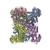
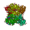
| Entry | Database: PDB / ID: 5xvv | ||||||
|---|---|---|---|---|---|---|---|
| Title | Crystal Structure of Forward Inhibited Aspergillus niger Glutamate Dehydrogenase With Both Apo- and Alpha Ketoglutarate Bound Subunits | ||||||
 Components Components | Glutamate dehydrogenase | ||||||
 Keywords Keywords | OXIDOREDUCTASE / Aspergillus / Glutamate / Dehydrogenase / 2-Oxoglutarate / Allostery / Forward Inhibition / 2-Mercaptoethanol | ||||||
| Function / homology |  Function and homology informationglutamate dehydrogenase (NADP+) activity / amino acid metabolic process / nucleotide binding Function and homology informationglutamate dehydrogenase (NADP+) activity / amino acid metabolic process / nucleotide bindingSimilarity search - Function | ||||||
| Biological species |  Aspergillus niger (mold) Aspergillus niger (mold) | ||||||
| Method | X-RAY DIFFRACTION / MOLECULAR REPLACEMENT / Resolution: 2.25 Å | ||||||
 Authors Authors | Prakash, P. / Punekar, N.S. / Bhaumik, P. | ||||||
| Funding support |  India, 1items India, 1items
| ||||||
 Citation Citation | Journal: J. Biol. Chem. / Year: 2018 Title: Structural basis for the catalytic mechanism and alpha-ketoglutarate cooperativity of glutamate dehydrogenase. Authors: Prakash, P. / Punekar, N.S. / Bhaumik, P. | ||||||
| History |
|
- Structure visualization
| Structure viewer | Molecule: MolmilJmol/JSmol |
|---|
- Downloads & links
-Download
| PDBx/mmCIF format | 5xvv.cif.gz | 536.9 KB | Display | PDBx/mmCIF format |
|---|---|---|---|---|
| PDB format | pdb5xvv.ent.gz | 459.3 KB | Display | PDB format |
| PDBx/mmJSON format | 5xvv.json.gz | Tree view | PDBx/mmJSON format | |
| Others |  Other downloads Other downloads |
-Validation report
| Arichive directory | https://data.pdbj.org/pub/pdb/validation_reports/xv/5xvvftp://data.pdbj.org/pub/pdb/validation_reports/xv/5xvv | HTTPS FTP |
|---|
-Related structure data


-Links
 PDBj
PDBj


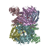
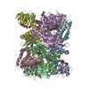
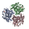
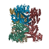
- Assembly
| Deposited unit | 
| ||||||||
|---|---|---|---|---|---|---|---|---|---|
| 1 |
| ||||||||
| Unit cell |
|
-Components
| #1: Protein | Mass: 49442.508 Da / Num. of mol.: 6 Source method: isolated from a genetically manipulated source Source: (gene. exp.) Aspergillus niger (mold)Production host: References: UniProt: B6V7E4 #2: Chemical | ChemComp-GOL / Glycerol  Mass: 92.094 Da / Num. of mol.: 22 / Source method: obtained synthetically / Formula: C3H8O3 Mass: 92.094 Da / Num. of mol.: 22 / Source method: obtained synthetically / Formula: C3H8O3#3: Chemical | Α-Ketoglutaric acid  Mass: 146.098 Da / Num. of mol.: 3 / Source method: obtained synthetically / Formula: C5H6O5 Mass: 146.098 Da / Num. of mol.: 3 / Source method: obtained synthetically / Formula: C5H6O5#4: Chemical | ChemComp-BME / 2-Mercaptoethanol  Mass: 78.133 Da / Num. of mol.: 6 / Source method: obtained synthetically / Formula: C2H6OS Mass: 78.133 Da / Num. of mol.: 6 / Source method: obtained synthetically / Formula: C2H6OS#5: Water | ChemComp-HOH / | Water Mass: 18.015 Da / Num. of mol.: 1516 / Source method: isolated from a natural source / Formula: H2O Mass: 18.015 Da / Num. of mol.: 1516 / Source method: isolated from a natural source / Formula: H2O |
|---|
-Experimental details
-Experiment
| Experiment | Method: X-RAY DIFFRACTION / Number of used crystals: 1 |
|---|
- Sample preparation
| Crystal | Density Matthews: 2.61 Å3/Da / Density % sol: 52.79 % |
|---|---|
| Crystal grow | Temperature: 295 K / Method: vapor diffusion, sitting drop / Details: 0.15 M potassium bromide, 20% w/v PEG 2000 MME |
-Data collection
| Diffraction | Mean temperature: 100 K |
|---|---|
| Diffraction source | Source: ROTATING ANODE / Type: RIGAKU MICROMAX-007 / Wavelength: 1.5418 Å |
| Detector | Type: RIGAKU RAXIS IV++ / Detector: IMAGE PLATE / Date: Oct 8, 2015 |
| Radiation | Protocol: SINGLE WAVELENGTH / Monochromatic (M) / Laue (L): M / Scattering type: x-ray |
| Radiation wavelength | Wavelength: 1.5418 Å / Relative weight: 1 |
| Reflection | Resolution: 2.25→35 Å / Num. obs: 135737 / % possible obs: 95.5 % / Redundancy: 2 % / CC1/2: 0.99 / Rmerge(I) obs: 0.095 / Net I/σ(I): 7.96 |
| Reflection shell | Resolution: 2.25→2.35 Å / Rmerge(I) obs: 0.39 / Mean I/σ(I) obs: 2.1 / Num. unique obs: 15991 / CC1/2: 0.68 / % possible all: 92 |
- Processing
| Software |
| ||||||||||||||||||||||||||||||||||||||||||||||||||||||||||||||||||||||||||||||||||||||||||||||||||||||||||||||||||||||||||||||||||||||||||||||||||||||||||||||||||||||||||||||||||||||
|---|---|---|---|---|---|---|---|---|---|---|---|---|---|---|---|---|---|---|---|---|---|---|---|---|---|---|---|---|---|---|---|---|---|---|---|---|---|---|---|---|---|---|---|---|---|---|---|---|---|---|---|---|---|---|---|---|---|---|---|---|---|---|---|---|---|---|---|---|---|---|---|---|---|---|---|---|---|---|---|---|---|---|---|---|---|---|---|---|---|---|---|---|---|---|---|---|---|---|---|---|---|---|---|---|---|---|---|---|---|---|---|---|---|---|---|---|---|---|---|---|---|---|---|---|---|---|---|---|---|---|---|---|---|---|---|---|---|---|---|---|---|---|---|---|---|---|---|---|---|---|---|---|---|---|---|---|---|---|---|---|---|---|---|---|---|---|---|---|---|---|---|---|---|---|---|---|---|---|---|---|---|---|---|
| Refinement | Method to determine structure: MOLECULAR REPLACEMENT / Resolution: 2.25→33 Å / Cor.coef. Fo:Fc: 0.961 / Cor.coef. Fo:Fc free: 0.937 / SU B: 6.523 / SU ML: 0.154 / Cross valid method: THROUGHOUT / ESU R: 0.282 / ESU R Free: 0.193 / Stereochemistry target values: MAXIMUM LIKELIHOOD / Details: HYDROGENS HAVE BEEN ADDED IN THE RIDING POSITIONS
| ||||||||||||||||||||||||||||||||||||||||||||||||||||||||||||||||||||||||||||||||||||||||||||||||||||||||||||||||||||||||||||||||||||||||||||||||||||||||||||||||||||||||||||||||||||||
| Solvent computation | Ion probe radii: 0.8 Å / Shrinkage radii: 0.8 Å / VDW probe radii: 1.2 Å / Solvent model: MASK | ||||||||||||||||||||||||||||||||||||||||||||||||||||||||||||||||||||||||||||||||||||||||||||||||||||||||||||||||||||||||||||||||||||||||||||||||||||||||||||||||||||||||||||||||||||||
| Displacement parameters | Biso mean: 25.927 Å2
| ||||||||||||||||||||||||||||||||||||||||||||||||||||||||||||||||||||||||||||||||||||||||||||||||||||||||||||||||||||||||||||||||||||||||||||||||||||||||||||||||||||||||||||||||||||||
| Refinement step | Cycle: 1 / Resolution: 2.25→33 Å
| ||||||||||||||||||||||||||||||||||||||||||||||||||||||||||||||||||||||||||||||||||||||||||||||||||||||||||||||||||||||||||||||||||||||||||||||||||||||||||||||||||||||||||||||||||||||
| Refine LS restraints |
|