 Movie
Movie Controller


+ Open data

- Basic information
| Entry | Database: PDB / ID: 5xv6 | ||||||
|---|---|---|---|---|---|---|---|
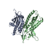
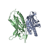
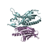
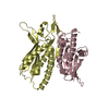
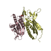
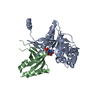
| Title | Crystal structure of ATG101-ATG13HORMA | ||||||
 Components Components |
| ||||||
 Keywords Keywords |  PROTEIN BINDING / AUTOPHAGY / ATG101 / ATG13 / HORMA PROTEIN BINDING / AUTOPHAGY / ATG101 / ATG13 / HORMA | ||||||
| Function / homology |  Function and homology informationregulation of protein lipidation / Atg1/ULK1 kinase complex / response to mitochondrial depolarisation / protein localization to phagophore assembly site / phagophore assembly site membrane / piecemeal microautophagy of the nucleus / protein kinase regulator activity / phagophore assembly site / positive regulation of protein targeting to mitochondrion / Macroautophagy ...regulation of protein lipidation / Atg1/ULK1 kinase complex / response to mitochondrial depolarisation / protein localization to phagophore assembly site / phagophore assembly site membrane / piecemeal microautophagy of the nucleus / protein kinase regulator activity / phagophore assembly site / positive regulation of protein targeting to mitochondrion / Macroautophagy / mitophagy / autophagosome assembly / autophagosome / positive regulation of autophagy / protein serine/threonine kinase activator activity / negative regulation of cell population proliferation / protein-containing complex binding / endoplasmic reticulum membrane / protein kinase binding / mitochondrion / identical protein binding / cytosol / cytoplasm Function and homology informationregulation of protein lipidation / Atg1/ULK1 kinase complex / response to mitochondrial depolarisation / protein localization to phagophore assembly site / phagophore assembly site membrane / piecemeal microautophagy of the nucleus / protein kinase regulator activity / phagophore assembly site / positive regulation of protein targeting to mitochondrion / Macroautophagy ...regulation of protein lipidation / Atg1/ULK1 kinase complex / response to mitochondrial depolarisation / protein localization to phagophore assembly site / phagophore assembly site membrane / piecemeal microautophagy of the nucleus / protein kinase regulator activity / phagophore assembly site / positive regulation of protein targeting to mitochondrion / Macroautophagy / mitophagy / autophagosome assembly / autophagosome / positive regulation of autophagy / protein serine/threonine kinase activator activity / negative regulation of cell population proliferation / protein-containing complex binding / endoplasmic reticulum membrane / protein kinase binding / mitochondrion / identical protein binding / cytosol / cytoplasmSimilarity search - Function | ||||||
| Biological species |  Homo sapiens (human) Homo sapiens (human) | ||||||
| Method | X-RAY DIFFRACTION / SYNCHROTRON / MOLECULAR REPLACEMENT / Resolution: 2.455 Å | ||||||
 Authors Authors | Kim, B.-W. / Song, H.K. | ||||||
 Citation Citation | Journal: Autophagy / Year: 2018 Title: The C-terminal region of ATG101 bridges ULK1 and PtdIns3K complex in autophagy initiation. Authors: Kim, B.-W. / Jin, Y. / Kim, J. / Kim, J.H. / Jung, J. / Kang, S. / Kim, I.Y. / Kim, J. / Cheong, H. / Song, H.K. | ||||||
| History |
|
- Structure visualization
| Structure viewer | Molecule: MolmilJmol/JSmol |
|---|
- Downloads & links
-Download
| PDBx/mmCIF format | 5xv6.cif.gz | 90.2 KB | Display | PDBx/mmCIF format |
|---|---|---|---|---|
| PDB format | pdb5xv6.ent.gz | 66.5 KB | Display | PDB format |
| PDBx/mmJSON format | 5xv6.json.gz | Tree view | PDBx/mmJSON format | |
| Others |  Other downloads Other downloads |
-Validation report
| Arichive directory | https://data.pdbj.org/pub/pdb/validation_reports/xv/5xv6ftp://data.pdbj.org/pub/pdb/validation_reports/xv/5xv6 | HTTPS FTP |
|---|
-Related structure data
| Related structure data |  5xuySC  5xv3C 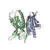 5xv4C S: Starting model for refinement C: citing same article ( |
|---|---|
| Similar structure data |


-Links
 PDBj
PDBj
- Assembly
| Deposited unit | 
| ||||||||
|---|---|---|---|---|---|---|---|---|---|
| 1 |
| ||||||||
| Unit cell |
|
-Components
| #1: Protein | Mass: 21319.652 Da / Num. of mol.: 1 / Fragment: UNP residues 1-190 Source method: isolated from a genetically manipulated source Source: (gene. exp.) Homo sapiens (human) / Gene: ATG13, KIAA0652 / Production host:  Escherichia coli (E. coli) / References: UniProt: O75143 Escherichia coli (E. coli) / References: UniProt: O75143 |
|---|---|
| #2: Protein | Mass: 24862.424 Da / Num. of mol.: 1 / Mutation: K40A/K41A/E42A Source method: isolated from a genetically manipulated source Source: (gene. exp.) Homo sapiens (human) / Gene: ATG101, C12orf44, PP894 / Production host: Escherichia coli (E. coli) / References: UniProt: Q9BSB4 |
-Experimental details
-Experiment
| Experiment | Method: X-RAY DIFFRACTION / Number of used crystals: 1 |
|---|
- Sample preparation
| Crystal | Density Matthews: 2.79 Å3/Da / Density % sol: 55.95 % |
|---|---|
| Crystal grow | Temperature: 293 K / Method: vapor diffusion, hanging drop Details: 0.1M HEPES (pH7.5), 0.2M MgCl2, 6-8% (w/v) PEG 3350 |
-Data collection
| Diffraction | Mean temperature: 100 K | |||||||||||||||||||||||||||||||||||||||||||||||||||||||||||||||||||||||||||||||||||||||||||||||||||||||||||||||||||||||||||||||||||||||||||||||||||||||||||||||||||||||||||||||||||||||||||||
|---|---|---|---|---|---|---|---|---|---|---|---|---|---|---|---|---|---|---|---|---|---|---|---|---|---|---|---|---|---|---|---|---|---|---|---|---|---|---|---|---|---|---|---|---|---|---|---|---|---|---|---|---|---|---|---|---|---|---|---|---|---|---|---|---|---|---|---|---|---|---|---|---|---|---|---|---|---|---|---|---|---|---|---|---|---|---|---|---|---|---|---|---|---|---|---|---|---|---|---|---|---|---|---|---|---|---|---|---|---|---|---|---|---|---|---|---|---|---|---|---|---|---|---|---|---|---|---|---|---|---|---|---|---|---|---|---|---|---|---|---|---|---|---|---|---|---|---|---|---|---|---|---|---|---|---|---|---|---|---|---|---|---|---|---|---|---|---|---|---|---|---|---|---|---|---|---|---|---|---|---|---|---|---|---|---|---|---|---|---|---|
| Diffraction source | Source: SYNCHROTRON / Site: Photon Factory  / Beamline: BL-17A / Wavelength: 0.98 Å / Beamline: BL-17A / Wavelength: 0.98 Å | |||||||||||||||||||||||||||||||||||||||||||||||||||||||||||||||||||||||||||||||||||||||||||||||||||||||||||||||||||||||||||||||||||||||||||||||||||||||||||||||||||||||||||||||||||||||||||||
| Detector | Type: DECTRIS PILATUS3 6M / Detector: PIXEL / Date: Dec 20, 2015 | |||||||||||||||||||||||||||||||||||||||||||||||||||||||||||||||||||||||||||||||||||||||||||||||||||||||||||||||||||||||||||||||||||||||||||||||||||||||||||||||||||||||||||||||||||||||||||||
| Radiation | Protocol: SINGLE WAVELENGTH / Monochromatic (M) / Laue (L): M / Scattering type: x-ray | |||||||||||||||||||||||||||||||||||||||||||||||||||||||||||||||||||||||||||||||||||||||||||||||||||||||||||||||||||||||||||||||||||||||||||||||||||||||||||||||||||||||||||||||||||||||||||||
| Radiation wavelength | Wavelength: 0.98 Å / Relative weight: 1 | |||||||||||||||||||||||||||||||||||||||||||||||||||||||||||||||||||||||||||||||||||||||||||||||||||||||||||||||||||||||||||||||||||||||||||||||||||||||||||||||||||||||||||||||||||||||||||||
| Reflection | Resolution: 2.44→50 Å / Num. obs: 19228 / % possible obs: 99.6 % / Redundancy: 7.1 % / Biso Wilson estimate: 64.06 Å2 / Rmerge(I) obs: 0.09 / Rpim(I) all: 0.038 / Rrim(I) all: 0.098 / Χ2: 1.575 / Net I/av σ(I): 33.39 / Net I/σ(I): 8.1 / Num. measured all: 136207 | |||||||||||||||||||||||||||||||||||||||||||||||||||||||||||||||||||||||||||||||||||||||||||||||||||||||||||||||||||||||||||||||||||||||||||||||||||||||||||||||||||||||||||||||||||||||||||||
| Reflection shell | Diffraction-ID: 1 / Rejects: 0
|
- Processing
| Software |
| |||||||||||||||||||||||||||||||||||||||||||||||||||||||||||||||||||||||||||||||||||||||||||||||||||||||||
|---|---|---|---|---|---|---|---|---|---|---|---|---|---|---|---|---|---|---|---|---|---|---|---|---|---|---|---|---|---|---|---|---|---|---|---|---|---|---|---|---|---|---|---|---|---|---|---|---|---|---|---|---|---|---|---|---|---|---|---|---|---|---|---|---|---|---|---|---|---|---|---|---|---|---|---|---|---|---|---|---|---|---|---|---|---|---|---|---|---|---|---|---|---|---|---|---|---|---|---|---|---|---|---|---|---|---|
| Refinement | Method to determine structure: MOLECULAR REPLACEMENT Starting model: 5XUY Resolution: 2.455→49.899 Å / SU ML: 0.35 / Cross valid method: FREE R-VALUE / σ(F): 1.36 / Phase error: 32.12
| |||||||||||||||||||||||||||||||||||||||||||||||||||||||||||||||||||||||||||||||||||||||||||||||||||||||||
| Solvent computation | Shrinkage radii: 0.9 Å / VDW probe radii: 1.11 Å | |||||||||||||||||||||||||||||||||||||||||||||||||||||||||||||||||||||||||||||||||||||||||||||||||||||||||
| Displacement parameters | Biso max: 147.23 Å2 / Biso mean: 77.5409 Å2 / Biso min: 38.17 Å2 | |||||||||||||||||||||||||||||||||||||||||||||||||||||||||||||||||||||||||||||||||||||||||||||||||||||||||
| Refinement step | Cycle: final / Resolution: 2.455→49.899 Å
| |||||||||||||||||||||||||||||||||||||||||||||||||||||||||||||||||||||||||||||||||||||||||||||||||||||||||
| Refine LS restraints |
| |||||||||||||||||||||||||||||||||||||||||||||||||||||||||||||||||||||||||||||||||||||||||||||||||||||||||
| LS refinement shell | Refine-ID: X-RAY DIFFRACTION / Total num. of bins used: 14
|
