 Movie
Movie Controller

[English] 日本語
 Yorodumi
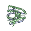
- PDB-5xub: The citrate-bound trimer of chemoreceptor MCP2201 ligand binding ... -
+ Open data

- Basic information
| Entry | Database: PDB / ID: 5xub | ||||||
|---|---|---|---|---|---|---|---|
| Title | The citrate-bound trimer of chemoreceptor MCP2201 ligand binding domain | ||||||
 Components Components | Methyl-accepting chemotaxis sensory transducer | ||||||
 Keywords Keywords |  SIGNALING PROTEIN / methyl-accepting chemotaxis protein / four helix bundle / dicarboxylic organic acid binding SIGNALING PROTEIN / methyl-accepting chemotaxis protein / four helix bundle / dicarboxylic organic acid binding | ||||||
| Function / homology |  Function and homology informationchemotaxis / transmembrane signaling receptor activity / signal transduction / membrane Function and homology informationchemotaxis / transmembrane signaling receptor activity / signal transduction / membraneSimilarity search - Function | ||||||
| Biological species |  Comamonas testosteroni (bacteria) Comamonas testosteroni (bacteria) | ||||||
| Method | X-RAY DIFFRACTION / SYNCHROTRON / MOLECULAR REPLACEMENT / Resolution: 2.5 Å | ||||||
 Authors Authors | Hong, Y. / Li, D.F. / Wang, D.C. | ||||||
 Citation Citation | Journal: Mol.Microbiol. / Year: 2019 Title: The ligand-binding domain of a chemoreceptor from Comamonas testosteroni has a previously unknown homotrimeric structure. Authors: Hong, Y. / Huang, Z. / Guo, L. / Ni, B. / Jiang, C.Y. / Li, X.J. / Hou, Y.J. / Yang, W.S. / Wang, D.C. / Zhulin, I.B. / Liu, S.J. / Li, D.F. | ||||||
| History |
|
- Structure visualization
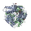
| Structure viewer | Molecule: MolmilJmol/JSmol |
|---|
- Downloads & links
-Download
| PDBx/mmCIF format | 5xub.cif.gz | 126.6 KB | Display | PDBx/mmCIF format |
|---|---|---|---|---|
| PDB format | pdb5xub.ent.gz | 98.8 KB | Display | PDB format |
| PDBx/mmJSON format | 5xub.json.gz | Tree view | PDBx/mmJSON format | |
| Others |  Other downloads Other downloads |
-Validation report
| Arichive directory | https://data.pdbj.org/pub/pdb/validation_reports/xu/5xubftp://data.pdbj.org/pub/pdb/validation_reports/xu/5xub | HTTPS FTP |
|---|
-Related structure data
| Related structure data | 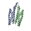 5xuaC  6itsC  5xud S: Starting model for refinement C: citing same article ( |
|---|---|
| Similar structure data |


-Links
 PDBj
PDBj

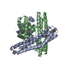
- Assembly
| Deposited unit | 
| |||||||||||||||
|---|---|---|---|---|---|---|---|---|---|---|---|---|---|---|---|---|
| 1 | 
| |||||||||||||||
| 2 | 
| |||||||||||||||
| Unit cell |
| |||||||||||||||
| Components on special symmetry positions |
|
-Components
| #1: Protein | Mass: 18138.564 Da / Num. of mol.: 2 / Fragment: UNP residues 46-203 Source method: isolated from a genetically manipulated source Source: (gene. exp.) Comamonas testosteroni (strain CNB-2) (bacteria)Strain: CNB-2 / Gene: CtCNB1_2201 / Production host: Escherichia coli BL21(DE3) (bacteria) / Strain (production host): BL21(DE3) / References: UniProt: D0IVL9#2: Chemical | Citric acid  Mass: 192.124 Da / Num. of mol.: 2 / Source method: obtained synthetically / Formula: C6H8O7 Mass: 192.124 Da / Num. of mol.: 2 / Source method: obtained synthetically / Formula: C6H8O7#3: Water | ChemComp-HOH / | Water Mass: 18.015 Da / Num. of mol.: 95 / Source method: isolated from a natural source / Formula: H2O Mass: 18.015 Da / Num. of mol.: 95 / Source method: isolated from a natural source / Formula: H2O |
|---|
-Experimental details
-Experiment
| Experiment | Method: X-RAY DIFFRACTION / Number of used crystals: 1 |
|---|
- Sample preparation
| Crystal | Density Matthews: 2.63 Å3/Da / Density % sol: 53.24 % |
|---|---|
| Crystal grow | Temperature: 295 K / Method: vapor diffusion, hanging drop Details: 0.1 M Sodium citrate pH 4.4, 14% PEG 400, 13% Ethylene glycol PH range: 4.0 - 4.8 |
-Data collection
| Diffraction | Mean temperature: 93 K |
|---|---|
| Diffraction source | Source: SYNCHROTRON / Site: SSRF  / Beamline: BL17U / Wavelength: 0.9789 Å / Beamline: BL17U / Wavelength: 0.9789 Å |
| Detector | Type: ADSC QUANTUM 315 / Detector: CCD / Date: Nov 26, 2013 |
| Radiation | Protocol: SINGLE WAVELENGTH / Monochromatic (M) / Laue (L): M / Scattering type: x-ray |
| Radiation wavelength | Wavelength: 0.9789 Å / Relative weight: 1 |
| Reflection | Resolution: 2.5→45.15 Å / Num. obs: 12095 / % possible obs: 94.5 % / Redundancy: 2.3 % / Biso Wilson estimate: 34.685 Å2 / CC1/2: 0.953 / Rmerge(I) obs: 0.152 / Net I/σ(I): 4.3 |
| Reflection shell | Resolution: 2.5→2.64 Å / Redundancy: 1.9 % / Rmerge(I) obs: 0.291 / Mean I/σ(I) obs: 2.2 / Num. unique obs: 1744 / CC1/2: 0.791 / % possible all: 94 |
- Processing
| Software |
| ||||||||||||||||||||||||||||||||||||||||||||||||||||||||||||||||||||||||||||||||||||||||||||||||||||||||||||||||||||||||||||||||||||||||||||||||||||||||||||||||||||||||||||||||||||||||||||||||||||||||||||||||||||||||||||||||||||||||||||||||||||||||||
|---|---|---|---|---|---|---|---|---|---|---|---|---|---|---|---|---|---|---|---|---|---|---|---|---|---|---|---|---|---|---|---|---|---|---|---|---|---|---|---|---|---|---|---|---|---|---|---|---|---|---|---|---|---|---|---|---|---|---|---|---|---|---|---|---|---|---|---|---|---|---|---|---|---|---|---|---|---|---|---|---|---|---|---|---|---|---|---|---|---|---|---|---|---|---|---|---|---|---|---|---|---|---|---|---|---|---|---|---|---|---|---|---|---|---|---|---|---|---|---|---|---|---|---|---|---|---|---|---|---|---|---|---|---|---|---|---|---|---|---|---|---|---|---|---|---|---|---|---|---|---|---|---|---|---|---|---|---|---|---|---|---|---|---|---|---|---|---|---|---|---|---|---|---|---|---|---|---|---|---|---|---|---|---|---|---|---|---|---|---|---|---|---|---|---|---|---|---|---|---|---|---|---|---|---|---|---|---|---|---|---|---|---|---|---|---|---|---|---|---|---|---|---|---|---|---|---|---|---|---|---|---|---|---|---|---|---|---|---|---|---|---|---|---|---|---|---|---|---|---|---|---|
| Refinement | Method to determine structure: MOLECULAR REPLACEMENT Starting model: 5XUD 5xud Resolution: 2.5→41.864 Å / SU ML: 0.32 / Cross valid method: FREE R-VALUE / σ(F): 1.97 / Phase error: 27.38
| ||||||||||||||||||||||||||||||||||||||||||||||||||||||||||||||||||||||||||||||||||||||||||||||||||||||||||||||||||||||||||||||||||||||||||||||||||||||||||||||||||||||||||||||||||||||||||||||||||||||||||||||||||||||||||||||||||||||||||||||||||||||||||
| Solvent computation | Shrinkage radii: 0.9 Å / VDW probe radii: 1.11 Å | ||||||||||||||||||||||||||||||||||||||||||||||||||||||||||||||||||||||||||||||||||||||||||||||||||||||||||||||||||||||||||||||||||||||||||||||||||||||||||||||||||||||||||||||||||||||||||||||||||||||||||||||||||||||||||||||||||||||||||||||||||||||||||
| Refinement step | Cycle: LAST / Resolution: 2.5→41.864 Å
| ||||||||||||||||||||||||||||||||||||||||||||||||||||||||||||||||||||||||||||||||||||||||||||||||||||||||||||||||||||||||||||||||||||||||||||||||||||||||||||||||||||||||||||||||||||||||||||||||||||||||||||||||||||||||||||||||||||||||||||||||||||||||||
| Refine LS restraints |
| ||||||||||||||||||||||||||||||||||||||||||||||||||||||||||||||||||||||||||||||||||||||||||||||||||||||||||||||||||||||||||||||||||||||||||||||||||||||||||||||||||||||||||||||||||||||||||||||||||||||||||||||||||||||||||||||||||||||||||||||||||||||||||
| LS refinement shell |
| ||||||||||||||||||||||||||||||||||||||||||||||||||||||||||||||||||||||||||||||||||||||||||||||||||||||||||||||||||||||||||||||||||||||||||||||||||||||||||||||||||||||||||||||||||||||||||||||||||||||||||||||||||||||||||||||||||||||||||||||||||||||||||
| Refinement TLS params. | Method: refined / Refine-ID: X-RAY DIFFRACTION
| ||||||||||||||||||||||||||||||||||||||||||||||||||||||||||||||||||||||||||||||||||||||||||||||||||||||||||||||||||||||||||||||||||||||||||||||||||||||||||||||||||||||||||||||||||||||||||||||||||||||||||||||||||||||||||||||||||||||||||||||||||||||||||
| Refinement TLS group |
|
