 Movie
Movie Controller

[English] 日本語
 Yorodumi
- PDB-5xoq: Crystal structure of O-Acetylserine Sulfhydrylase with bound Tran... -
+ Open data

- Basic information
| Entry | Database: PDB / ID: 5xoq | ||||||
|---|---|---|---|---|---|---|---|
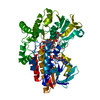
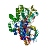
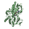
| Title | Crystal structure of O-Acetylserine Sulfhydrylase with bound Transcription Factor peptide inhibitor from Planctomyces limnophilus | ||||||
 Components Components |
| ||||||
 Keywords Keywords | TRANSFERASE/TRANSFERASE INHIBITOR / CysK /  OASS / Transcription Factor / Tf peptide / Cysteine Synthase / Planctomyces limnophilus / TRANSFERASE-TRANSFERASE INHIBITOR complex OASS / Transcription Factor / Tf peptide / Cysteine Synthase / Planctomyces limnophilus / TRANSFERASE-TRANSFERASE INHIBITOR complex | ||||||
| Function / homology |  Function and homology informationcysteine synthase / cysteine synthase activity / cysteine biosynthetic process from serine Function and homology informationcysteine synthase / cysteine synthase activity / cysteine biosynthetic process from serineSimilarity search - Function | ||||||
| Biological species | Planctopirus limnophila Planctomyces limnophilus (bacteria) Planctomyces limnophilus (bacteria) | ||||||
| Method | X-RAY DIFFRACTION / MOLECULAR REPLACEMENT / Resolution: 1.87 Å | ||||||
 Authors Authors | Singh, R.P. / Saini, N. | ||||||
| Funding support |  India, 1items India, 1items
| ||||||
 Citation Citation | Journal: To Be Published Title: Crystal structure of O-Acetylserine Sulfhydrylase with bound Transcription Factor peptide inhibitor from Planctomyces limnophilus Authors: Singh, R.P. / Saini, N. #1: Journal: To Be PublishedTitle: Crystal structure of O-acetylserine sulfhydrylase with bound peptide of C-term transcription factor as inhibitor from planctomyces limnophilus Authors: Singh, R.P. / Saini, N. | ||||||
| History |
|
- Structure visualization
| Structure viewer | Molecule: MolmilJmol/JSmol |
|---|
- Downloads & links
-Download
| PDBx/mmCIF format | 5xoq.cif.gz | 139 KB | Display | PDBx/mmCIF format |
|---|---|---|---|---|
| PDB format | pdb5xoq.ent.gz | 107.1 KB | Display | PDB format |
| PDBx/mmJSON format | 5xoq.json.gz | Tree view | PDBx/mmJSON format | |
| Others |  Other downloads Other downloads |
-Validation report
| Arichive directory | https://data.pdbj.org/pub/pdb/validation_reports/xo/5xoqftp://data.pdbj.org/pub/pdb/validation_reports/xo/5xoq | HTTPS FTP |
|---|
-Related structure data
| Related structure data | 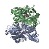 5xa2S S: Starting model for refinement |
|---|---|
| Similar structure data |


-Links
 PDBj
PDBj
- Assembly
| Deposited unit | 
| ||||||||
|---|---|---|---|---|---|---|---|---|---|
| 1 |
| ||||||||
| Unit cell |
|
-Components
| #1: Protein | / O-Acetylserine Sulfhydrylase Mass: 33022.883 Da / Num. of mol.: 2 Source method: isolated from a genetically manipulated source Source: (gene. exp.) Planctopirus limnophila (strain ATCC 43296 / DSM 3776 / IFAM 1008 / 290) (bacteria)Strain: ATCC 43296 / DSM 3776 / IFAM 1008 / 290 / Gene: Plim_3256 / Production host: Escherichia coli (E. coli) / Strain (production host): BL21(DE3) / References: UniProt: D5STP0, cysteine synthase#2: Protein/peptide | Mass: 708.720 Da / Num. of mol.: 2 / Source method: obtained synthetically / Source: (synth.) Planctomyces limnophilus (bacteria)#3: Chemical | Diethylene glycol  Mass: 106.120 Da / Num. of mol.: 2 / Source method: obtained synthetically / Formula: C4H10O3 Mass: 106.120 Da / Num. of mol.: 2 / Source method: obtained synthetically / Formula: C4H10O3#4: Water | ChemComp-HOH / | Water Mass: 18.015 Da / Num. of mol.: 393 / Source method: isolated from a natural source / Formula: H2O Mass: 18.015 Da / Num. of mol.: 393 / Source method: isolated from a natural source / Formula: H2O |
|---|
-Experimental details
-Experiment
| Experiment | Method: X-RAY DIFFRACTION / Number of used crystals: 1 |
|---|
- Sample preparation
| Crystal | Density Matthews: 2.62 Å3/Da / Density % sol: 53.02 % |
|---|---|
| Crystal grow | Temperature: 291.15 K / Method: vapor diffusion, sitting drop / pH: 8.5 / Details: 20% PEG 8000, 100mM Tris pH8.5, 200mM MgCl2 |
-Data collection
| Diffraction | Mean temperature: 100 K |
|---|---|
| Diffraction source | Source: SEALED TUBE / Type: RIGAKU MICROMAX-002 / Wavelength: 1.54 Å |
| Detector | Type: MAR scanner 345 mm plate / Detector: IMAGE PLATE / Date: May 19, 2016 |
| Radiation | Protocol: SINGLE WAVELENGTH / Monochromatic (M) / Laue (L): M / Scattering type: x-ray |
| Radiation wavelength | Wavelength: 1.54 Å / Relative weight: 1 |
| Reflection | Resolution: 1.87→25.22 Å / Num. obs: 54061 / % possible obs: 93 % / Redundancy: 1.1 % / Biso Wilson estimate: 18.27 Å2 / CC1/2: 1 / Rmerge(I) obs: 0.055 / Χ2: 0.956 / Net I/av σ(I): 22.6 / Net I/σ(I): 15.6 |
| Reflection shell | Resolution: 1.87→1.9 Å / Redundancy: 4 % / Rmerge(I) obs: 0.202 / Mean I/σ(I) obs: 4.61 / Num. unique obs: 2483 / CC1/2: 1 / Χ2: 0.7 / % possible all: 86.2 |
- Processing
| Software |
| |||||||||||||||||||||||||||||||||||||||||||||||||||||||||||||||||||||||||||||||||||||||||||||||||||||||||||||||||||||||||||||||||||||||||||||||||||
|---|---|---|---|---|---|---|---|---|---|---|---|---|---|---|---|---|---|---|---|---|---|---|---|---|---|---|---|---|---|---|---|---|---|---|---|---|---|---|---|---|---|---|---|---|---|---|---|---|---|---|---|---|---|---|---|---|---|---|---|---|---|---|---|---|---|---|---|---|---|---|---|---|---|---|---|---|---|---|---|---|---|---|---|---|---|---|---|---|---|---|---|---|---|---|---|---|---|---|---|---|---|---|---|---|---|---|---|---|---|---|---|---|---|---|---|---|---|---|---|---|---|---|---|---|---|---|---|---|---|---|---|---|---|---|---|---|---|---|---|---|---|---|---|---|---|---|---|---|
| Refinement | Method to determine structure: MOLECULAR REPLACEMENT Starting model: 5XA2 Resolution: 1.87→25.216 Å / SU ML: 0.17 / Cross valid method: FREE R-VALUE / σ(F): 1.34 / Phase error: 18.24 / Stereochemistry target values: ML
| |||||||||||||||||||||||||||||||||||||||||||||||||||||||||||||||||||||||||||||||||||||||||||||||||||||||||||||||||||||||||||||||||||||||||||||||||||
| Solvent computation | Shrinkage radii: 0.9 Å / VDW probe radii: 1.11 Å / Solvent model: FLAT BULK SOLVENT MODEL | |||||||||||||||||||||||||||||||||||||||||||||||||||||||||||||||||||||||||||||||||||||||||||||||||||||||||||||||||||||||||||||||||||||||||||||||||||
| Refinement step | Cycle: LAST / Resolution: 1.87→25.216 Å
| |||||||||||||||||||||||||||||||||||||||||||||||||||||||||||||||||||||||||||||||||||||||||||||||||||||||||||||||||||||||||||||||||||||||||||||||||||
| Refine LS restraints |
| |||||||||||||||||||||||||||||||||||||||||||||||||||||||||||||||||||||||||||||||||||||||||||||||||||||||||||||||||||||||||||||||||||||||||||||||||||
| LS refinement shell |
|
