 Movie
Movie Controller

[English] 日本語
 Yorodumi
- PDB-5xmh: Crystal structure of an IgM rheumatoid factor YES8c in complex wi... -
+ Open data

- Basic information
| Entry | Database: PDB / ID: 5xmh | |||||||||
|---|---|---|---|---|---|---|---|---|---|---|
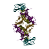
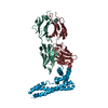
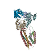
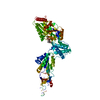
| Title | Crystal structure of an IgM rheumatoid factor YES8c in complex with IgG1 Fc | |||||||||
 Components Components |
| |||||||||
 Keywords Keywords | IMMUNE SYSTEM / Immunoglobulin / Complex / Rheumatoid factor / Autoantibody | |||||||||
| Function / homology |  Function and homology information Function and homology informationimmunoglobulin complex, circulating / immunoglobulin receptor binding / complement activation, classical pathway / antigen binding / antibacterial humoral response / blood microparticle / extracellular exosome / plasma membrane Similarity search - Function | |||||||||
| Biological species |  Homo sapiens (human) Homo sapiens (human) | |||||||||
| Method |  X-RAY DIFFRACTION / SYNCHROTRON / Resolution: 2.8 Å X-RAY DIFFRACTION / SYNCHROTRON / Resolution: 2.8 Å | |||||||||
 Authors Authors | Shiroishi, M. / Shimokawa, K. / Lee, J.M. / Kusakabe, M. / Ueda, T. | |||||||||
| Funding support |  Japan, 2items Japan, 2items
| |||||||||
 Citation Citation | Journal: J. Biol. Chem. / Year: 2018 Title: Structure-function analyses of a stereotypic rheumatoid factor unravel the structural basis for germline-encoded antibody autoreactivity. Authors: Shiroishi, M. / Ito, Y. / Shimokawa, K. / Lee, J.M. / Kusakabe, T. / Ueda, T. | |||||||||
| History |
|
- Structure visualization
| Structure viewer | Molecule: MolmilJmol/JSmol |
|---|
- Downloads & links
-Download
| PDBx/mmCIF format | 5xmh.cif.gz | 217.1 KB | Display | PDBx/mmCIF format |
|---|---|---|---|---|
| PDB format | pdb5xmh.ent.gz | 172.3 KB | Display | PDB format |
| PDBx/mmJSON format | 5xmh.json.gz | Tree view | PDBx/mmJSON format | |
| Others |  Other downloads Other downloads |
-Validation report
| Summary document | 5xmh_validation.pdf.gz | 1.2 MB | Display | wwPDB validaton report |
|---|---|---|---|---|
| Full document | 5xmh_full_validation.pdf.gz | 1.2 MB | Display | |
| Data in XML | 5xmh_validation.xml.gz | 38 KB | Display | |
| Data in CIF | 5xmh_validation.cif.gz | 52.7 KB | Display | |
| Arichive directory | https://data.pdbj.org/pub/pdb/validation_reports/xm/5xmhftp://data.pdbj.org/pub/pdb/validation_reports/xm/5xmh | HTTPS FTP |
-Related structure data
| Similar structure data |
|---|


-Links
 PDBj
PDBj


- Assembly
| Deposited unit | 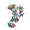
| ||||||||
|---|---|---|---|---|---|---|---|---|---|
| 1 | 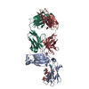
| ||||||||
| 2 | 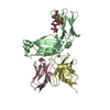
| ||||||||
| Unit cell |
|
-Components
-Antibody , 3 types, 6 molecules ABLCHD
| #1: Antibody | Mass: 23634.676 Da / Num. of mol.: 2 / Fragment: UNP RESIDUES 239-446 Source method: isolated from a genetically manipulated source Source: (gene. exp.) Homo sapiens (human) / Production host:   Cricetulus griseus (Chinese hamster) / References: UniProt: P0DOX5 Cricetulus griseus (Chinese hamster) / References: UniProt: P0DOX5#2: Antibody | Mass: 23625.207 Da / Num. of mol.: 2 Source method: isolated from a genetically manipulated source Source: (gene. exp.) Homo sapiens (human) / Plasmid: pET22b / Production host:  #3: Antibody | Mass: 23799.732 Da / Num. of mol.: 2 Source method: isolated from a genetically manipulated source Source: (gene. exp.) Homo sapiens (human) / Plasmid: pET22b / Production host: |
|---|
-Sugars , 2 types, 2 molecules
| #4: Polysaccharide | 2-acetamido-2-deoxy-beta-D-glucopyranose-(1-2)-alpha-D-mannopyranose-(1-3)-[2-acetamido-2-deoxy- ...2-acetamido-2-deoxy-beta-D-glucopyranose-(1-2)-alpha-D-mannopyranose-(1-3)-[2-acetamido-2-deoxy-beta-D-glucopyranose-(1-2)-alpha-D-mannopyranose-(1-6)]beta-D-mannopyranose-(1-4)-2-acetamido-2-deoxy-beta-D-glucopyranose-(1-4)-[alpha-L-fucopyranose-(1-6)]2-acetamido-2-deoxy-beta-D-glucopyranose Source method: isolated from a genetically manipulated source |
|---|---|
| #5: Polysaccharide | 2-acetamido-2-deoxy-beta-D-glucopyranose-(1-2)-alpha-D-mannopyranose-(1-6)-[alpha-D-mannopyranose- ...2-acetamido-2-deoxy-beta-D-glucopyranose-(1-2)-alpha-D-mannopyranose-(1-6)-[alpha-D-mannopyranose-(1-3)]beta-D-mannopyranose-(1-4)-2-acetamido-2-deoxy-beta-D-glucopyranose-(1-4)-2-acetamido-2-deoxy-beta-D-glucopyranose Source method: isolated from a genetically manipulated source |
-Non-polymers , 1 types, 64 molecules 
| #6: Water | ChemComp-HOH / Mass: 18.015 Da / Num. of mol.: 64 / Source method: isolated from a natural source / Formula: H2O |
|---|
-Experimental details
-Experiment
| Experiment | Method: X-RAY DIFFRACTION / Number of used crystals: 1 |
|---|
- Sample preparation
| Crystal | Density Matthews: 2.92 Å3/Da / Density % sol: 57.81 % Description: The entry contains friedel pairs in F_plus/minus columns and I_plus/minus columns |
|---|---|
| Crystal grow | Temperature: 293 K / Method: vapor diffusion, sitting drop / pH: 8.5 / Details: Tris-HCl, sodium acetate, PEG 4000 |
-Data collection
| Diffraction | Mean temperature: 100 K |
|---|---|
| Diffraction source | Source: SYNCHROTRON / Site: Photon Factory / Beamline: AR-NE3A / Wavelength: 1 Å |
| Detector | Type: DECTRIS PILATUS 2M-F / Detector: PIXEL / Date: Jun 15, 2015 |
| Radiation | Protocol: SINGLE WAVELENGTH / Monochromatic (M) / Laue (L): M / Scattering type: x-ray |
| Radiation wavelength | Wavelength: 1 Å / Relative weight: 1 |
| Reflection | Resolution: 2.8→20 Å / Num. obs: 77576 / % possible obs: 98.7 % / Redundancy: 4.6 % / Rmerge(I) obs: 0.074 / Net I/σ(I): 19.1 |
| Reflection shell | Resolution: 2.8→2.85 Å / Redundancy: 3.8 % / Rmerge(I) obs: 0.355 / Num. unique obs: 3825 / CC1/2: 0.917 / % possible all: 95.1 |
- Processing
| Software |
| ||||||||||||||||||||
|---|---|---|---|---|---|---|---|---|---|---|---|---|---|---|---|---|---|---|---|---|---|
| Refinement | Resolution: 2.8→19.9 Å / Cross valid method: FREE R-VALUE Details: The entry contains friedel pairs in F_plus/minus columns and I_plus/minus columns
| ||||||||||||||||||||
| Displacement parameters | Biso mean: 52 Å2 | ||||||||||||||||||||
| Refinement step | Cycle: LAST / Resolution: 2.8→19.9 Å
|
