 Movie
Movie Controller


+ Open data

- Basic information
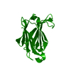
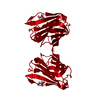
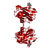
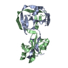
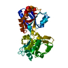
| Entry | Database: PDB / ID: 5xg8 | ||||||
|---|---|---|---|---|---|---|---|
| Title | Galectin-13/Placental Protein 13 variant R53H crystal structure | ||||||
 Components Components | Galactoside-binding soluble lectin 13 | ||||||
 Keywords Keywords | SUGAR BINDING PROTEIN /  disulfide bond disulfide bond | ||||||
| Function / homology |  Function and homology informationlysophospholipase activity / phospholipid metabolic process / nuclear matrix / carbohydrate binding / nuclear body / apoptotic process / nucleoplasm / cytoplasm Function and homology informationlysophospholipase activity / phospholipid metabolic process / nuclear matrix / carbohydrate binding / nuclear body / apoptotic process / nucleoplasm / cytoplasmSimilarity search - Function | ||||||
| Biological species |  Homo sapiens (human) Homo sapiens (human) | ||||||
| Method | X-RAY DIFFRACTION / SYNCHROTRON / MOLECULAR REPLACEMENT / Resolution: 1.55 Å | ||||||
 Authors Authors | Wang, Y. / Su, J.Y. | ||||||
| Funding support |  China, 1items China, 1items
| ||||||
 Citation Citation | Journal: Sci Rep / Year: 2018 Title: Galectin-13, a different prototype galectin, does not bind beta-galacto-sides and forms dimers via intermolecular disulfide bridges between Cys-136 and Cys-138 Authors: Su, J. / Wang, Y. / Si, Y. / Gao, J. / Song, C. / Cui, L. / Wu, R. / Tai, G. / Zhou, Y. | ||||||
| History |
|
- Structure visualization
| Structure viewer | Molecule: MolmilJmol/JSmol |
|---|
- Downloads & links
-Download
| PDBx/mmCIF format | 5xg8.cif.gz | 64.4 KB | Display | PDBx/mmCIF format |
|---|---|---|---|---|
| PDB format | pdb5xg8.ent.gz | 51.4 KB | Display | PDB format |
| PDBx/mmJSON format | 5xg8.json.gz | Tree view | PDBx/mmJSON format | |
| Others |  Other downloads Other downloads |
-Validation report
| Arichive directory | https://data.pdbj.org/pub/pdb/validation_reports/xg/5xg8ftp://data.pdbj.org/pub/pdb/validation_reports/xg/5xg8 | HTTPS FTP |
|---|
-Related structure data


-Links
 PDBj
PDBj- Assembly
| Deposited unit | 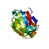
| ||||||||||||
|---|---|---|---|---|---|---|---|---|---|---|---|---|---|
| 1 | 
| ||||||||||||
| Unit cell |
| ||||||||||||
| Components on special symmetry positions |
|
-Components
| #1: Protein | Mass: 15985.277 Da / Num. of mol.: 1 / Fragment: UNP residues 2-139 / Mutation: R53H Source method: isolated from a genetically manipulated source Source: (gene. exp.) Homo sapiens (human) / Gene: LGALS13, PLAC8 / Production host:  Escherichia coli (E. coli) / References: UniProt: Q9UHV8 Escherichia coli (E. coli) / References: UniProt: Q9UHV8 |
|---|---|
| #2: Chemical | ChemComp-GOL / Glycerol  Mass: 92.094 Da / Num. of mol.: 1 / Source method: obtained synthetically / Formula: C3H8O3 Mass: 92.094 Da / Num. of mol.: 1 / Source method: obtained synthetically / Formula: C3H8O3 |
| #3: Water | ChemComp-HOH / Water Mass: 18.015 Da / Num. of mol.: 124 / Source method: isolated from a natural source / Formula: H2O Mass: 18.015 Da / Num. of mol.: 124 / Source method: isolated from a natural source / Formula: H2O |
-Experimental details
-Experiment
| Experiment | Method: X-RAY DIFFRACTION / Number of used crystals: 1 |
|---|
- Sample preparation
| Crystal | Density Matthews: 2.12 Å3/Da / Density % sol: 41.93 % |
|---|---|
| Crystal grow | Temperature: 298 K / Method: vapor diffusion, hanging drop / Details: PEG |
-Data collection
| Diffraction | Mean temperature: 100 K |
|---|---|
| Diffraction source | Source: SYNCHROTRON / Site: SSRF / Beamline: BL18U1 / Wavelength: 0.9 Å |
| Detector | Type: DECTRIS PILATUS3 6M / Detector: PIXEL / Date: Mar 19, 2017 |
| Radiation | Protocol: SINGLE WAVELENGTH / Monochromatic (M) / Laue (L): M / Scattering type: x-ray |
| Radiation wavelength | Wavelength: 0.9 Å / Relative weight: 1 |
| Reflection | Resolution: 1.55→19.092 Å / Num. obs: 20107 / % possible obs: 99.5 % / Redundancy: 6.2 % / Net I/σ(I): 18.3 |
- Processing
| Software |
| ||||||||||||||||||||||||
|---|---|---|---|---|---|---|---|---|---|---|---|---|---|---|---|---|---|---|---|---|---|---|---|---|---|
| Refinement | Method to determine structure: MOLECULAR REPLACEMENT / Resolution: 1.55→19.092 Å / SU ML: 0.13 / Cross valid method: NONE / σ(F): 1.35 / Phase error: 25.44 / Stereochemistry target values: ML
| ||||||||||||||||||||||||
| Solvent computation | Shrinkage radii: 0.9 Å / VDW probe radii: 1.11 Å / Solvent model: FLAT BULK SOLVENT MODEL | ||||||||||||||||||||||||
| Displacement parameters | Biso max: 95.82 Å2 / Biso mean: 30.9956 Å2 / Biso min: 17.12 Å2 | ||||||||||||||||||||||||
| Refinement step | Cycle: final / Resolution: 1.55→19.092 Å
|
