 Movie
Movie Controller

[English] 日本語
 Yorodumi
- PDB-3k6u: M. acetivorans Molybdate-Binding Protein (ModA) in Unliganded Ope... -
+ Open data

- Basic information
| Entry | Database: PDB / ID: 3k6u | ||||||
|---|---|---|---|---|---|---|---|
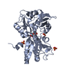
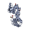
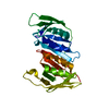
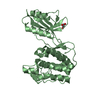
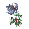
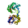
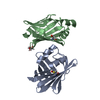
| Title | M. acetivorans Molybdate-Binding Protein (ModA) in Unliganded Open Form | ||||||
 Components Components | Solute-binding protein MA_0280 | ||||||
 Keywords Keywords |  TRANSPORT PROTEIN / ModA / molybdate / Methanosarcina acetivorans / periplasmic binding protein / ABC transporter / ligand / metal-binding protein TRANSPORT PROTEIN / ModA / molybdate / Methanosarcina acetivorans / periplasmic binding protein / ABC transporter / ligand / metal-binding protein | ||||||
| Function / homology | Tungstate ABC transporter, substrate-binding protein WtpA / tungstate binding / Bacterial extracellular solute-binding protein / Periplasmic binding protein-like II / D-Maltodextrin-Binding Protein; domain 2 / 3-Layer(aba) Sandwich / Alpha Beta / Uncharacterized solute-binding protein MA_0280 Function and homology information Function and homology information | ||||||
| Biological species |  Methanosarcina acetivorans (archaea) Methanosarcina acetivorans (archaea) | ||||||
| Method | X-RAY DIFFRACTION / SYNCHROTRON / MIRAS / Resolution: 1.95 Å | ||||||
 Authors Authors | Chan, S. / Giuroiu, I. / Chernishof, I. / Sawaya, M.R. / Chiang, J. / Gunsalus, R.P. / Arbing, M.A. / Perry, L.J. | ||||||
 Citation Citation | Journal: Acta Crystallogr.,Sect.F / Year: 2010 Title: Apo and ligand-bound structures of ModA from the archaeon Methanosarcina acetivorans Authors: Chan, S. / Giuroiu, I. / Chernishof, I. / Sawaya, M.R. / Chiang, J. / Gunsalus, R.P. / Arbing, M.A. / Perry, L.J. | ||||||
| History |
|
- Structure visualization
| Structure viewer | Molecule: MolmilJmol/JSmol |
|---|
- Downloads & links
-Download
| PDBx/mmCIF format | 3k6u.cif.gz | 79.6 KB | Display | PDBx/mmCIF format |
|---|---|---|---|---|
| PDB format | pdb3k6u.ent.gz | 57.5 KB | Display | PDB format |
| PDBx/mmJSON format | 3k6u.json.gz | Tree view | PDBx/mmJSON format | |
| Others |  Other downloads Other downloads |
-Validation report
| Arichive directory | https://data.pdbj.org/pub/pdb/validation_reports/k6/3k6uftp://data.pdbj.org/pub/pdb/validation_reports/k6/3k6u | HTTPS FTP |
|---|
-Related structure data


-Links
 PDBj
PDBj- Assembly
| Deposited unit | 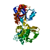
| ||||||||
|---|---|---|---|---|---|---|---|---|---|
| 1 |
| ||||||||
| Unit cell |
|
-Components
| #1: Protein | Mass: 39160.156 Da / Num. of mol.: 1 / Fragment: UNP residues 27 to 352 Source method: isolated from a genetically manipulated source Details: N-terminal TEV-cleavable 6xHis-tag / Source: (gene. exp.) Methanosarcina acetivorans (archaea) / Strain: C2A / Gene: MA0280, MA_0280 / Plasmid: pETM-11 / Production host:  Escherichia coli (E. coli) / Strain (production host): BL21-Gold (DE3) / References: UniProt: Q8TTZ5 Escherichia coli (E. coli) / Strain (production host): BL21-Gold (DE3) / References: UniProt: Q8TTZ5 |
|---|---|
| #2: Water | ChemComp-HOH / Water Mass: 18.015 Da / Num. of mol.: 176 / Source method: isolated from a natural source / Formula: H2O Mass: 18.015 Da / Num. of mol.: 176 / Source method: isolated from a natural source / Formula: H2O |
-Experimental details
-Experiment
| Experiment | Method: X-RAY DIFFRACTION / Number of used crystals: 1 |
|---|
- Sample preparation
| Crystal |
| ||||||||||||||||||||
|---|---|---|---|---|---|---|---|---|---|---|---|---|---|---|---|---|---|---|---|---|---|
| Crystal grow |
|
-Data collection
| Diffraction |
| ||||||||||||||||||||||||||||||||||||||||||||||||||||||||||||||||||
|---|---|---|---|---|---|---|---|---|---|---|---|---|---|---|---|---|---|---|---|---|---|---|---|---|---|---|---|---|---|---|---|---|---|---|---|---|---|---|---|---|---|---|---|---|---|---|---|---|---|---|---|---|---|---|---|---|---|---|---|---|---|---|---|---|---|---|---|
| Diffraction source |
| ||||||||||||||||||||||||||||||||||||||||||||||||||||||||||||||||||
| Detector |
| ||||||||||||||||||||||||||||||||||||||||||||||||||||||||||||||||||
| Radiation |
| ||||||||||||||||||||||||||||||||||||||||||||||||||||||||||||||||||
| Radiation wavelength |
| ||||||||||||||||||||||||||||||||||||||||||||||||||||||||||||||||||
| Reflection | Av σ(I) over netI: 22.45 / Number: 195952 / Rmerge(I) obs: 0.124 / Χ2: 1 / D res high: 2.7 Å / D res low: 90 Å / Num. obs: 21339 / % possible obs: 100 | ||||||||||||||||||||||||||||||||||||||||||||||||||||||||||||||||||
| Diffraction reflection shell |
| ||||||||||||||||||||||||||||||||||||||||||||||||||||||||||||||||||
| Reflection | Resolution: 1.94→38.26 Å / Num. obs: 29447 / % possible obs: 99.9 % / Redundancy: 10.86 % / Rmerge(I) obs: 0.092 / Rsym value: 0.079 / Χ2: 1.03 / Net I/σ(I): 18.6 | ||||||||||||||||||||||||||||||||||||||||||||||||||||||||||||||||||
| Reflection shell | Resolution: 1.94→2.01 Å / Rmerge(I) obs: 0.5 / Mean I/σ(I) obs: 10.43 / Num. unique all: 2936 / Rsym value: 0.468 / Χ2: 1.021 / % possible all: 100 |

-Phasing
| Phasing | Method: MIRAS | |||||||||||||||||||||||||||||||||||||||||||||||||||||||||||||||||||||||||||||||||||||||||||||||||||||||||||||||||||||||||||||||||||||||||||||||||||||||||||||||||||||||||||||||||||||||||||
|---|---|---|---|---|---|---|---|---|---|---|---|---|---|---|---|---|---|---|---|---|---|---|---|---|---|---|---|---|---|---|---|---|---|---|---|---|---|---|---|---|---|---|---|---|---|---|---|---|---|---|---|---|---|---|---|---|---|---|---|---|---|---|---|---|---|---|---|---|---|---|---|---|---|---|---|---|---|---|---|---|---|---|---|---|---|---|---|---|---|---|---|---|---|---|---|---|---|---|---|---|---|---|---|---|---|---|---|---|---|---|---|---|---|---|---|---|---|---|---|---|---|---|---|---|---|---|---|---|---|---|---|---|---|---|---|---|---|---|---|---|---|---|---|---|---|---|---|---|---|---|---|---|---|---|---|---|---|---|---|---|---|---|---|---|---|---|---|---|---|---|---|---|---|---|---|---|---|---|---|---|---|---|---|---|---|---|---|---|
| Phasing dm | Method: Solvent flattening and Histogram matching / Reflection: 29027 | |||||||||||||||||||||||||||||||||||||||||||||||||||||||||||||||||||||||||||||||||||||||||||||||||||||||||||||||||||||||||||||||||||||||||||||||||||||||||||||||||||||||||||||||||||||||||||
| Phasing dm shell |
| |||||||||||||||||||||||||||||||||||||||||||||||||||||||||||||||||||||||||||||||||||||||||||||||||||||||||||||||||||||||||||||||||||||||||||||||||||||||||||||||||||||||||||||||||||||||||||
| Phasing MIR | Resolution: 1.95→20 Å / FOM: 0.368 / FOM acentric: 0.363 / FOM centric: 0.513 / Reflection: 10848 / Reflection acentric: 10438 / Reflection centric: 410 | |||||||||||||||||||||||||||||||||||||||||||||||||||||||||||||||||||||||||||||||||||||||||||||||||||||||||||||||||||||||||||||||||||||||||||||||||||||||||||||||||||||||||||||||||||||||||||
| Phasing MIR der | Der set-ID: 1
| |||||||||||||||||||||||||||||||||||||||||||||||||||||||||||||||||||||||||||||||||||||||||||||||||||||||||||||||||||||||||||||||||||||||||||||||||||||||||||||||||||||||||||||||||||||||||||
| Phasing MIR der shell |
| |||||||||||||||||||||||||||||||||||||||||||||||||||||||||||||||||||||||||||||||||||||||||||||||||||||||||||||||||||||||||||||||||||||||||||||||||||||||||||||||||||||||||||||||||||||||||||
| Phasing MIR der site |
| |||||||||||||||||||||||||||||||||||||||||||||||||||||||||||||||||||||||||||||||||||||||||||||||||||||||||||||||||||||||||||||||||||||||||||||||||||||||||||||||||||||||||||||||||||||||||||
| Phasing MIR shell |
|
- Processing
| Software |
| |||||||||||||||||||||||||||||||||||||||||||||||||||||||||||||||||
|---|---|---|---|---|---|---|---|---|---|---|---|---|---|---|---|---|---|---|---|---|---|---|---|---|---|---|---|---|---|---|---|---|---|---|---|---|---|---|---|---|---|---|---|---|---|---|---|---|---|---|---|---|---|---|---|---|---|---|---|---|---|---|---|---|---|---|
| Refinement | Method to determine structure: MIRAS / Resolution: 1.95→38.26 Å / Cor.coef. Fo:Fc: 0.96 / Cor.coef. Fo:Fc free: 0.947 / WRfactor Rfree: 0.234 / WRfactor Rwork: 0.197 / Occupancy max: 1 / Occupancy min: 0 / FOM work R set: 0.87 / SU B: 6.561 / SU ML: 0.086 / SU R Cruickshank DPI: 0.136 / SU Rfree: 0.127 / TLS residual ADP flag: LIKELY RESIDUAL / Cross valid method: THROUGHOUT / σ(F): 0 / ESU R: 0.136 / ESU R Free: 0.127 / Stereochemistry target values: MAXIMUM LIKELIHOOD / Details: U VALUES: RESIDUAL ONLY
| |||||||||||||||||||||||||||||||||||||||||||||||||||||||||||||||||
| Solvent computation | Ion probe radii: 0.8 Å / Shrinkage radii: 0.8 Å / VDW probe radii: 1.4 Å / Solvent model: MASK | |||||||||||||||||||||||||||||||||||||||||||||||||||||||||||||||||
| Displacement parameters | Biso max: 54.9 Å2 / Biso mean: 17.613 Å2 / Biso min: 5.61 Å2
| |||||||||||||||||||||||||||||||||||||||||||||||||||||||||||||||||
| Refinement step | Cycle: LAST / Resolution: 1.95→38.26 Å
| |||||||||||||||||||||||||||||||||||||||||||||||||||||||||||||||||
| Refine LS restraints |
| |||||||||||||||||||||||||||||||||||||||||||||||||||||||||||||||||
| LS refinement shell | Resolution: 1.95→2.001 Å / Total num. of bins used: 20
| |||||||||||||||||||||||||||||||||||||||||||||||||||||||||||||||||
| Refinement TLS params. | Method: refined / Origin x: 15.8757 Å / Origin y: 25.8208 Å / Origin z: 14.2498 Å
| |||||||||||||||||||||||||||||||||||||||||||||||||||||||||||||||||
| Refinement TLS group |
|
