 Movie
Movie Controller

[English] 日本語
 Yorodumi
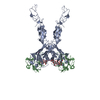
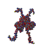
- PDB-5xg3: Crystal structure of the ATPgS-engaged Smc head domain with an ex... -
+ Open data

- Basic information
| Entry | Database: PDB / ID: 5xg3 | ||||||
|---|---|---|---|---|---|---|---|
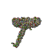
| Title | Crystal structure of the ATPgS-engaged Smc head domain with an extended coiled coil bound to the C-terminal domain of ScpA derived from Bacillus subtilis | ||||||
 Components Components |
| ||||||
 Keywords Keywords | DNA BINDING PROTEIN/CELL CYCLE /  Condensin / Smc / ATPase / ScpA / DNA BINDING PROTEIN-CELL CYCLE complex Condensin / Smc / ATPase / ScpA / DNA BINDING PROTEIN-CELL CYCLE complex | ||||||
| Function / homology |  Function and homology information Function and homology informationchromosome condensation / sister chromatid cohesion / chromosome segregation / chromosome / DNA replication / cell division / ATP hydrolysis activity / DNA binding / ATP binding / identical protein binding / cytoplasmSimilarity search - Function | ||||||
| Biological species |  Bacillus subtilis (bacteria) Bacillus subtilis (bacteria) | ||||||
| Method | X-RAY DIFFRACTION / SYNCHROTRON / MOLECULAR REPLACEMENT / Resolution: 3.5 Å | ||||||
 Authors Authors | Shin, H.-C. / Lee, H. / Oh, B.-H. | ||||||
 Citation Citation | Journal: Mol. Cell / Year: 2017 Title: Structure of Full-Length SMC and Rearrangements Required for Chromosome Organization Authors: Diebold-Durand, M.L. / Lee, H. / Ruiz Avila, L.B. / Noh, H. / Shin, H.C. / Im, H. / Bock, F.P. / Burmann, F. / Durand, A. / Basfeld, A. / Ham, S. / Basquin, J. / Oh, B.-H. / Gruber, S. | ||||||
| History |
|
- Structure visualization
| Structure viewer | Molecule: MolmilJmol/JSmol |
|---|
- Downloads & links
-Download
| PDBx/mmCIF format | 5xg3.cif.gz | 173.4 KB | Display | PDBx/mmCIF format |
|---|---|---|---|---|
| PDB format | pdb5xg3.ent.gz | 124.9 KB | Display | PDB format |
| PDBx/mmJSON format | 5xg3.json.gz | Tree view | PDBx/mmJSON format | |
| Others |  Other downloads Other downloads |
-Validation report
| Arichive directory | https://data.pdbj.org/pub/pdb/validation_reports/xg/5xg3ftp://data.pdbj.org/pub/pdb/validation_reports/xg/5xg3 | HTTPS FTP |
|---|
-Related structure data
| Related structure data |  5nmoC  5nnvC 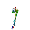 5xeiC 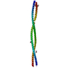 5xg2C 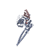 5xnsC  1xexS C: citing same article ( S: Starting model for refinement |
|---|---|
| Similar structure data |


-Links
 PDBj
PDBj

- Assembly
| Deposited unit | 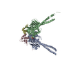
| ||||||||
|---|---|---|---|---|---|---|---|---|---|
| 1 |
| ||||||||
| Unit cell |
|
-Components
-Protein , 2 types, 4 molecules ABCD
| #1: Protein | Mass: 49153.883 Da / Num. of mol.: 2 / Fragment: UNP residues 1-219,UNP residues 975-1186 / Mutation: E1118Q,E1118Q Source method: isolated from a genetically manipulated source Source: (gene. exp.) Bacillus subtilis (strain 168) (bacteria)Strain: 168 / Gene: smc, ylqA, BSU15940 / Production host: Escherichia coli (E. coli) / References: UniProt: P51834#2: Protein | Mass: 10367.890 Da / Num. of mol.: 2 / Fragment: UNP residues 167-251 Source method: isolated from a genetically manipulated source Source: (gene. exp.) Bacillus subtilis (bacteria) / Gene: scpA, SAMN05878487_2386 / Production host: Escherichia coli (E. coli) / References: UniProt: A0A1N6WAJ8, UniProt: P35154*PLUS |
|---|
-Non-polymers , 4 types, 8 molecules 


| #3: Chemical |  Mass: 523.247 Da / Num. of mol.: 2 / Source method: obtained synthetically / Formula: C10H16N5O12P3S / Comment: ATP-gamma-S, energy-carrying molecule analogue*YM Mass: 523.247 Da / Num. of mol.: 2 / Source method: obtained synthetically / Formula: C10H16N5O12P3S / Comment: ATP-gamma-S, energy-carrying molecule analogue*YM#4: Chemical |  Mass: 24.305 Da / Num. of mol.: 2 / Source method: obtained synthetically / Formula: Mg Mass: 24.305 Da / Num. of mol.: 2 / Source method: obtained synthetically / Formula: Mg#5: Chemical | ChemComp-CO / |  Mass: 58.933 Da / Num. of mol.: 1 / Source method: obtained synthetically / Formula: Co Mass: 58.933 Da / Num. of mol.: 1 / Source method: obtained synthetically / Formula: Co#6: Water | ChemComp-HOH / | WaterMass: 18.015 Da / Num. of mol.: 3 / Source method: isolated from a natural source / Formula: H2O |
|---|
-Experimental details
-Experiment
| Experiment | Method: X-RAY DIFFRACTION / Number of used crystals: 1 |
|---|
- Sample preparation
| Crystal | Density Matthews: 3.59 Å3/Da / Density % sol: 65.75 % |
|---|---|
| Crystal grow | Temperature: 295 K / Method: vapor diffusion, hanging drop / pH: 8 Details: 10% PEG 3000, 0.1M Imidazole pH8.0, 0.2M LiSO4, 0.05M Hexamine cobalt (III) chloride |
-Data collection
| Diffraction | Mean temperature: 100 K |
|---|---|
| Diffraction source | Source: SYNCHROTRON / Site: Photon Factory  / Beamline: BL-5A / Wavelength: 1 Å / Beamline: BL-5A / Wavelength: 1 Å |
| Detector | Type: ADSC QUANTUM 315 / Detector: CCD / Date: Jun 20, 2014 |
| Radiation | Protocol: SINGLE WAVELENGTH / Monochromatic (M) / Laue (L): M / Scattering type: x-ray |
| Radiation wavelength | Wavelength: 1 Å / Relative weight: 1 |
| Reflection | Resolution: 3→50 Å / Num. obs: 35074 / % possible obs: 92.6 % / Redundancy: 5.4 % / Biso Wilson estimate: 57.51 Å2 / Net I/σ(I): 12.3 |
- Processing
| Software |
| ||||||||||||||||||||||||||||||||||||||||||||||||||||||||||||||||||||||||||||||||||||||||||||||||||||||||||||||||||||||||||||||
|---|---|---|---|---|---|---|---|---|---|---|---|---|---|---|---|---|---|---|---|---|---|---|---|---|---|---|---|---|---|---|---|---|---|---|---|---|---|---|---|---|---|---|---|---|---|---|---|---|---|---|---|---|---|---|---|---|---|---|---|---|---|---|---|---|---|---|---|---|---|---|---|---|---|---|---|---|---|---|---|---|---|---|---|---|---|---|---|---|---|---|---|---|---|---|---|---|---|---|---|---|---|---|---|---|---|---|---|---|---|---|---|---|---|---|---|---|---|---|---|---|---|---|---|---|---|---|---|
| Refinement | Method to determine structure: MOLECULAR REPLACEMENT Starting model: 1XEX Resolution: 3.5→40.989 Å / SU ML: 0.5 / Cross valid method: FREE R-VALUE / σ(F): 1.7 / Phase error: 27.08 / Stereochemistry target values: ML
| ||||||||||||||||||||||||||||||||||||||||||||||||||||||||||||||||||||||||||||||||||||||||||||||||||||||||||||||||||||||||||||||
| Solvent computation | Shrinkage radii: 0.9 Å / VDW probe radii: 1.11 Å / Solvent model: FLAT BULK SOLVENT MODEL | ||||||||||||||||||||||||||||||||||||||||||||||||||||||||||||||||||||||||||||||||||||||||||||||||||||||||||||||||||||||||||||||
| Displacement parameters | Biso max: 149.68 Å2 / Biso mean: 54.4176 Å2 / Biso min: 3.91 Å2 | ||||||||||||||||||||||||||||||||||||||||||||||||||||||||||||||||||||||||||||||||||||||||||||||||||||||||||||||||||||||||||||||
| Refinement step | Cycle: final / Resolution: 3.5→40.989 Å
| ||||||||||||||||||||||||||||||||||||||||||||||||||||||||||||||||||||||||||||||||||||||||||||||||||||||||||||||||||||||||||||||
| Refine LS restraints |
| ||||||||||||||||||||||||||||||||||||||||||||||||||||||||||||||||||||||||||||||||||||||||||||||||||||||||||||||||||||||||||||||
| LS refinement shell | Refine-ID: X-RAY DIFFRACTION / Rfactor Rfree error: 0 / Total num. of bins used: 17
|
