 Movie
Movie Controller

[English] 日本語
 Yorodumi
- PDB-5x5m: Crystal structure of a hydrolase encoded by lin2189 from Listeria... -
+ Open data

- Basic information
| Entry | Database: PDB / ID: 5x5m | ||||||
|---|---|---|---|---|---|---|---|
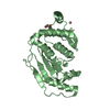
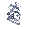
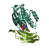
| Title | Crystal structure of a hydrolase encoded by lin2189 from Listeria innocua | ||||||
 Components Components | Lin2189 protein | ||||||
 Keywords Keywords | HYDROLASE/ANTIBIOTIC /  hydrolase / HYDROLASE-ANTIBIOTIC complex hydrolase / HYDROLASE-ANTIBIOTIC complex | ||||||
| Function / homology |  Function and homology information Function and homology informationGyrI-like cyclopropanoid cyclopropyl hydrolase Lin2189-like / GyrI-like small molecule binding domain / Multidrug-efflux Transporter 1 Regulator Bmrr; Chain A / Regulatory factor, effector binding domain / GyrI-like small molecule binding domain / Regulatory factor, effector binding domain superfamily / Alpha-Beta Barrel / Alpha Beta Similarity search - Domain/homology | ||||||
| Biological species | Listeria innocua serovar 6a | ||||||
| Method | X-RAY DIFFRACTION / SYNCHROTRON / MOLECULAR REPLACEMENT / Resolution: 1.211 Å | ||||||
 Authors Authors | Zhang, J. | ||||||
 Citation Citation | Journal: Nat Commun / Year: 2017 Title: GyrI-like proteins catalyze cyclopropanoid hydrolysis to confer cellular protection Authors: Yuan, H. / Zhang, J. / Cai, Y. / Wu, S. / Yang, K. / Chan, H.C.S. / Huang, W. / Jin, W.B. / Li, Y. / Yin, Y. / Igarashi, Y. / Yuan, S. / Zhou, J. / Tang, G.L. | ||||||
| History |
|
- Structure visualization
| Structure viewer | Molecule: MolmilJmol/JSmol |
|---|
- Downloads & links
-Download
| PDBx/mmCIF format | 5x5m.cif.gz | 111.5 KB | Display | PDBx/mmCIF format |
|---|---|---|---|---|
| PDB format | pdb5x5m.ent.gz | 84.2 KB | Display | PDB format |
| PDBx/mmJSON format | 5x5m.json.gz | Tree view | PDBx/mmJSON format | |
| Others |  Other downloads Other downloads |
-Validation report
| Arichive directory | https://data.pdbj.org/pub/pdb/validation_reports/x5/5x5mftp://data.pdbj.org/pub/pdb/validation_reports/x5/5x5m | HTTPS FTP |
|---|
-Related structure data
| Related structure data | 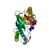 3b49S S: Starting model for refinement |
|---|---|
| Similar structure data |


-Links
 PDBj
PDBj- Assembly
| Deposited unit | 
| |||||||||
|---|---|---|---|---|---|---|---|---|---|---|
| 1 | 
| |||||||||
| 2 | 
| |||||||||
| Unit cell |
| |||||||||
| Components on special symmetry positions |
|
-Components
| #1: Protein | Mass: 25363.141 Da / Num. of mol.: 2 / Mutation: E157A/E185L Source method: isolated from a genetically manipulated source Source: (gene. exp.)  Listeria innocua serovar 6a (strain ATCC BAA-680 / CLIP 11262) (bacteria) Listeria innocua serovar 6a (strain ATCC BAA-680 / CLIP 11262) (bacteria)Strain: ATCC BAA-680 / CLIP 11262 / Gene: lin2189 / Production host:  Enterobacteria phage L1 (virus) / References: UniProt: Q929T5 Enterobacteria phage L1 (virus) / References: UniProt: Q929T5#2: Chemical | Dimethyl sulfoxide  Mass: 78.133 Da / Num. of mol.: 2 Mass: 78.133 Da / Num. of mol.: 2Source method: isolated from a genetically manipulated source Formula: C2H6OS / Comment: DMSO, precipitant*YM #3: Chemical |   Mass: 679.698 Da / Num. of mol.: 2 / Source method: isolated from a natural source / Formula: C35H29N5O8S Mass: 679.698 Da / Num. of mol.: 2 / Source method: isolated from a natural source / Formula: C35H29N5O8S#4: Water | ChemComp-HOH / | Water Mass: 18.015 Da / Num. of mol.: 428 / Source method: isolated from a natural source / Formula: H2O Mass: 18.015 Da / Num. of mol.: 428 / Source method: isolated from a natural source / Formula: H2O |
|---|
-Experimental details
-Experiment
| Experiment | Method: X-RAY DIFFRACTION / Number of used crystals: 1 |
|---|
- Sample preparation
| Crystal | Density Matthews: 2.02 Å3/Da / Density % sol: 39.01 % |
|---|---|
| Crystal grow | Temperature: 289 K / Method: vapor diffusion, sitting drop / pH: 6.5 / Details: PEG 4000, Glycerol, MES/imidazole |
-Data collection
| Diffraction | Mean temperature: 100 K |
|---|---|
| Diffraction source | Source: SYNCHROTRON / Site: SSRF  / Beamline: BL19U1 / Wavelength: 0.97791 Å / Beamline: BL19U1 / Wavelength: 0.97791 Å |
| Detector | Type: DECTRIS PILATUS3 S 6M / Detector: PIXEL / Date: Dec 12, 2016 |
| Radiation | Protocol: SINGLE WAVELENGTH / Monochromatic (M) / Laue (L): M / Scattering type: x-ray |
| Radiation wavelength | Wavelength: 0.97791 Å / Relative weight: 1 |
| Reflection | Resolution: 1.21→50 Å / Num. obs: 112031 / % possible obs: 96 % / Redundancy: 6.9 % / Rmerge(I) obs: 0.061 / Rpim(I) all: 0.025 / Net I/σ(I): 30.5 |
- Processing
| Software |
| |||||||||||||||||||||||||||||||||||||||||||||||||||||||||||||||||||||||||||||||||||||||||||||||||||||||||||||||||||||||||||||||||||||||||||||||||||||||||||||||||||||||||||||||||||||||||||||||||||||||||||||||||||||||||
|---|---|---|---|---|---|---|---|---|---|---|---|---|---|---|---|---|---|---|---|---|---|---|---|---|---|---|---|---|---|---|---|---|---|---|---|---|---|---|---|---|---|---|---|---|---|---|---|---|---|---|---|---|---|---|---|---|---|---|---|---|---|---|---|---|---|---|---|---|---|---|---|---|---|---|---|---|---|---|---|---|---|---|---|---|---|---|---|---|---|---|---|---|---|---|---|---|---|---|---|---|---|---|---|---|---|---|---|---|---|---|---|---|---|---|---|---|---|---|---|---|---|---|---|---|---|---|---|---|---|---|---|---|---|---|---|---|---|---|---|---|---|---|---|---|---|---|---|---|---|---|---|---|---|---|---|---|---|---|---|---|---|---|---|---|---|---|---|---|---|---|---|---|---|---|---|---|---|---|---|---|---|---|---|---|---|---|---|---|---|---|---|---|---|---|---|---|---|---|---|---|---|---|---|---|---|---|---|---|---|---|---|---|---|---|---|---|---|---|
| Refinement | Method to determine structure: MOLECULAR REPLACEMENT Starting model: 3B49 Resolution: 1.211→44.039 Å / SU ML: 0.11 / Cross valid method: FREE R-VALUE / σ(F): 1.34 / Phase error: 19 / Stereochemistry target values: ML
| |||||||||||||||||||||||||||||||||||||||||||||||||||||||||||||||||||||||||||||||||||||||||||||||||||||||||||||||||||||||||||||||||||||||||||||||||||||||||||||||||||||||||||||||||||||||||||||||||||||||||||||||||||||||||
| Solvent computation | Shrinkage radii: 0.9 Å / VDW probe radii: 1.11 Å / Solvent model: FLAT BULK SOLVENT MODEL | |||||||||||||||||||||||||||||||||||||||||||||||||||||||||||||||||||||||||||||||||||||||||||||||||||||||||||||||||||||||||||||||||||||||||||||||||||||||||||||||||||||||||||||||||||||||||||||||||||||||||||||||||||||||||
| Refinement step | Cycle: LAST / Resolution: 1.211→44.039 Å
| |||||||||||||||||||||||||||||||||||||||||||||||||||||||||||||||||||||||||||||||||||||||||||||||||||||||||||||||||||||||||||||||||||||||||||||||||||||||||||||||||||||||||||||||||||||||||||||||||||||||||||||||||||||||||
| Refine LS restraints |
| |||||||||||||||||||||||||||||||||||||||||||||||||||||||||||||||||||||||||||||||||||||||||||||||||||||||||||||||||||||||||||||||||||||||||||||||||||||||||||||||||||||||||||||||||||||||||||||||||||||||||||||||||||||||||
| LS refinement shell |
|
