 Movie
Movie Controller

[English] 日本語
 Yorodumi
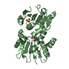
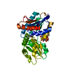
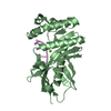
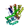
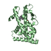
- PDB-2iwi: CRYSTAL STRUCTURE OF THE HUMAN PIM2 IN COMPLEX WITH A RUTHENIUM O... -
+ Open data

- Basic information
| Entry | Database: PDB / ID: 2iwi | ||||||
|---|---|---|---|---|---|---|---|
| Title | CRYSTAL STRUCTURE OF THE HUMAN PIM2 IN COMPLEX WITH A RUTHENIUM ORGANOMETALLIC LIGAND RU1 | ||||||
 Components Components | SERINE/THREONINE-PROTEIN KINASE PIM-2 | ||||||
 Keywords Keywords |  TRANSFERASE / NUCLEOTIDE-BINDING / SERINE/THREONINE-PROTEIN KINASE / PIM2 / KINASE / CANCER / LEUKEMIA / ATP-BINDING / PROTO-ONCOGENE / PHOSPHORYLATION TRANSFERASE / NUCLEOTIDE-BINDING / SERINE/THREONINE-PROTEIN KINASE / PIM2 / KINASE / CANCER / LEUKEMIA / ATP-BINDING / PROTO-ONCOGENE / PHOSPHORYLATION | ||||||
| Function / homology |  Function and homology information Function and homology informationapoptotic mitochondrial changes / positive regulation of macroautophagy / positive regulation of autophagy / regulation of mitotic cell cycle / macroautophagy / response to virus / G1/S transition of mitotic cell cycle / positive regulation of canonical NF-kappaB signal transduction / protein stabilization / non-specific serine/threonine protein kinase ...apoptotic mitochondrial changes / positive regulation of macroautophagy / positive regulation of autophagy / regulation of mitotic cell cycle / macroautophagy / response to virus / G1/S transition of mitotic cell cycle / positive regulation of canonical NF-kappaB signal transduction / protein stabilization / non-specific serine/threonine protein kinase / negative regulation of cell population proliferation / protein phosphorylation / protein serine kinase activity / protein serine/threonine kinase activity / negative regulation of apoptotic process / positive regulation of DNA-templated transcription / ATP binding / cytoplasmSimilarity search - Function | ||||||
| Biological species |  HOMO SAPIENS (human) HOMO SAPIENS (human) | ||||||
| Method | X-RAY DIFFRACTION / SYNCHROTRON / MOLECULAR REPLACEMENT / Resolution: 2.8 Å | ||||||
 Authors Authors | Russo, S. / Debreczeni, J.E. / Amos, A. / Bullock, A.N. / Fedorov, O. / Niesen, F. / Sobott, F. / Turnbull, A. / Pike, A.C.W. / Ugochukwu, E. ...Russo, S. / Debreczeni, J.E. / Amos, A. / Bullock, A.N. / Fedorov, O. / Niesen, F. / Sobott, F. / Turnbull, A. / Pike, A.C.W. / Ugochukwu, E. / Papagrigoriou, E. / Bunkoczi, G. / Gorrec, F. / Edwards, A. / Arrowsmith, C. / Weigelt, J. / Sundstrom, M. / von Delft, F. / Knapp, S. | ||||||
 Citation Citation | Journal: PLoS ONE / Year: 2009 Title: Crystal structure of the PIM2 kinase in complex with an organoruthenium inhibitor. Authors: Bullock, A.N. / Russo, S. / Amos, A. / Pagano, N. / Bregman, H. / Debreczeni, J.E. / Lee, W.H. / von Delft, F. / Meggers, E. / Knapp, S. | ||||||
| History |
|
- Structure visualization
| Structure viewer | Molecule: MolmilJmol/JSmol |
|---|
- Downloads & links
-Download
| PDBx/mmCIF format | 2iwi.cif.gz | 107.6 KB | Display | PDBx/mmCIF format |
|---|---|---|---|---|
| PDB format | pdb2iwi.ent.gz | 81 KB | Display | PDB format |
| PDBx/mmJSON format | 2iwi.json.gz | Tree view | PDBx/mmJSON format | |
| Others |  Other downloads Other downloads |
-Validation report
| Arichive directory | https://data.pdbj.org/pub/pdb/validation_reports/iw/2iwiftp://data.pdbj.org/pub/pdb/validation_reports/iw/2iwi | HTTPS FTP |
|---|
-Related structure data
| Related structure data |  1xwsS S: Starting model for refinement |
|---|---|
| Similar structure data |


-Links
 PDBj
PDBj
- Assembly
| Deposited unit | 
| ||||||||||||||||||||||||||||||||||||||||||||||||
|---|---|---|---|---|---|---|---|---|---|---|---|---|---|---|---|---|---|---|---|---|---|---|---|---|---|---|---|---|---|---|---|---|---|---|---|---|---|---|---|---|---|---|---|---|---|---|---|---|---|
| 1 | 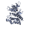
| ||||||||||||||||||||||||||||||||||||||||||||||||
| 2 | 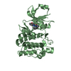
| ||||||||||||||||||||||||||||||||||||||||||||||||
| Unit cell |
| ||||||||||||||||||||||||||||||||||||||||||||||||
| Noncrystallographic symmetry (NCS) | NCS domain:
NCS domain segments:
NCS ensembles :
NCS oper: (Code: given Matrix: (0.94987, 0.28264, -0.13363), Vector : |
-Components
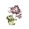
| #1: Protein | Mass: 34316.375 Da / Num. of mol.: 2 Source method: isolated from a genetically manipulated source Source: (gene. exp.) HOMO SAPIENS (human) / Production host:  Escherichia coli BL21(DE3) (bacteria) / Variant (production host): Star Escherichia coli BL21(DE3) (bacteria) / Variant (production host): StarReferences: UniProt: Q9P1W9, non-specific serine/threonine protein kinase#2: Chemical | 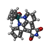  Mass: 480.438 Da / Num. of mol.: 2 / Source method: obtained synthetically / Formula: C23H13N3O3Ru Mass: 480.438 Da / Num. of mol.: 2 / Source method: obtained synthetically / Formula: C23H13N3O3Ru |
|---|
-Experimental details
-Experiment
| Experiment | Method: X-RAY DIFFRACTION / Number of used crystals: 1 |
|---|
- Sample preparation
| Crystal | Density Matthews: 2.63 Å3/Da / Density % sol: 53.18 % |
|---|---|
| Crystal grow | Method: vapor diffusion, sitting drop Details: 1.5 UL SITTING DROPS, 1.6 M SODIUM-POTASSIUM PHOSPHATE, 0.1 M HEPES PH 7.5 |
-Data collection
| Diffraction | Mean temperature: 100 K |
|---|---|
| Diffraction source | Source: SYNCHROTRON / Site: SLS  / Beamline: X10SA / Wavelength: 0.9789 / Beamline: X10SA / Wavelength: 0.9789 |
| Detector | Type: MARRESEARCH / Detector: CCD / Date: Mar 4, 2006 / Details: MIRRORS |
| Radiation | Monochromator: SI 111 / Protocol: SINGLE WAVELENGTH / Monochromatic (M) / Laue (L): M / Scattering type: x-ray |
| Radiation wavelength | Wavelength: 0.9789 Å / Relative weight: 1 |
| Reflection | Resolution: 2.8→44.19 Å / Num. obs: 16854 / % possible obs: 99.7 % / Observed criterion σ(I): 2 / Redundancy: 2.9 % / Rmerge(I) obs: 0.06 / Net I/σ(I): 11.23 |
| Reflection shell | Resolution: 2.8→2.9 Å / Redundancy: 2.9 % / Rmerge(I) obs: 0.38 / Mean I/σ(I) obs: 2.21 / % possible all: 100 |
- Processing
| Software |
| ||||||||||||||||||||||||||||||||||||||||||||||||||||||||||||||||||||||||||||||||||||||||||||||||||||||||||||||||||||||||||||||||||||||||||||||||||||||||||||||||||||||||||||||||||||||
|---|---|---|---|---|---|---|---|---|---|---|---|---|---|---|---|---|---|---|---|---|---|---|---|---|---|---|---|---|---|---|---|---|---|---|---|---|---|---|---|---|---|---|---|---|---|---|---|---|---|---|---|---|---|---|---|---|---|---|---|---|---|---|---|---|---|---|---|---|---|---|---|---|---|---|---|---|---|---|---|---|---|---|---|---|---|---|---|---|---|---|---|---|---|---|---|---|---|---|---|---|---|---|---|---|---|---|---|---|---|---|---|---|---|---|---|---|---|---|---|---|---|---|---|---|---|---|---|---|---|---|---|---|---|---|---|---|---|---|---|---|---|---|---|---|---|---|---|---|---|---|---|---|---|---|---|---|---|---|---|---|---|---|---|---|---|---|---|---|---|---|---|---|---|---|---|---|---|---|---|---|---|---|---|
| Refinement | Method to determine structure: MOLECULAR REPLACEMENT Starting model: PDB ENTRY 1XWS Resolution: 2.8→76.47 Å / Cor.coef. Fo:Fc: 0.929 / Cor.coef. Fo:Fc free: 0.89 / SU B: 47.383 / SU ML: 0.408 / Cross valid method: THROUGHOUT / ESU R: 1.485 / ESU R Free: 0.402 / Stereochemistry target values: MAXIMUM LIKELIHOOD / Details: HYDROGENS HAVE BEEN ADDED IN THE RIDING POSITIONS.
| ||||||||||||||||||||||||||||||||||||||||||||||||||||||||||||||||||||||||||||||||||||||||||||||||||||||||||||||||||||||||||||||||||||||||||||||||||||||||||||||||||||||||||||||||||||||
| Solvent computation | Ion probe radii: 0.8 Å / Shrinkage radii: 0.8 Å / VDW probe radii: 1.4 Å / Solvent model: MASK | ||||||||||||||||||||||||||||||||||||||||||||||||||||||||||||||||||||||||||||||||||||||||||||||||||||||||||||||||||||||||||||||||||||||||||||||||||||||||||||||||||||||||||||||||||||||
| Displacement parameters | Biso mean: 20.47 Å2
| ||||||||||||||||||||||||||||||||||||||||||||||||||||||||||||||||||||||||||||||||||||||||||||||||||||||||||||||||||||||||||||||||||||||||||||||||||||||||||||||||||||||||||||||||||||||
| Refinement step | Cycle: LAST / Resolution: 2.8→76.47 Å
| ||||||||||||||||||||||||||||||||||||||||||||||||||||||||||||||||||||||||||||||||||||||||||||||||||||||||||||||||||||||||||||||||||||||||||||||||||||||||||||||||||||||||||||||||||||||
| Refine LS restraints |
|