 Movie
Movie Controller


+ Open data

- Basic information
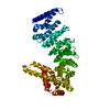
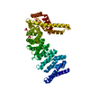
| Entry | Database: PDB / ID: 5wqt | ||||||
|---|---|---|---|---|---|---|---|
| Title | Structure of a protein involved in pyroptosis | ||||||
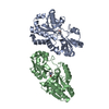
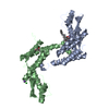
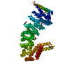
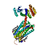
 Components Components | Gasdermin-D | ||||||
 Keywords Keywords |  SIGNALING PROTEIN / pyrtosis SIGNALING PROTEIN / pyrtosis | ||||||
| Function / homology |  Function and homology information Function and homology informationpore complex assembly / : / Release of apoptotic factors from the mitochondria / wide pore channel activity / NLRP3 inflammasome complex / cardiolipin binding / Regulation of TLR by endogenous ligand / phosphatidic acid binding / Interleukin-1 processing / phosphatidylinositol-4-phosphate binding ...pore complex assembly / : / Release of apoptotic factors from the mitochondria / wide pore channel activity / NLRP3 inflammasome complex / cardiolipin binding / Regulation of TLR by endogenous ligand / phosphatidic acid binding / Interleukin-1 processing / phosphatidylinositol-4-phosphate binding / phosphatidylserine binding / pyroptotic inflammatory response / protein secretion / Pyroptosis / Purinergic signaling in leishmaniasis infection / phosphatidylinositol-4,5-bisphosphate binding / positive regulation of interleukin-1 beta production / mitochondrial membrane / protein homooligomerization / positive regulation of inflammatory response / specific granule lumen / tertiary granule lumen / ficolin-1-rich granule lumen / defense response to Gram-negative bacterium / defense response to Gram-positive bacterium / defense response to bacterium / inflammatory response / innate immune response / Neutrophil degranulation / extracellular space / extracellular region / nucleoplasm / membrane / plasma membrane / cytosolSimilarity search - Function | ||||||
| Biological species |  Homo sapiens (human) Homo sapiens (human) | ||||||
| Method | X-RAY DIFFRACTION / SYNCHROTRON / MOLECULAR REPLACEMENT / Resolution: 2.64 Å | ||||||
 Authors Authors | Kuang, S. / Li, J. | ||||||
 Citation Citation | Journal: Proc. Natl. Acad. Sci. U.S.A. / Year: 2017 Title: Structure insight of GSDMD reveals the basis of GSDMD autoinhibition in cell pyroptosis. Authors: Kuang, S. / Zheng, J. / Yang, H. / Li, S. / Duan, S. / Shen, Y. / Ji, C. / Gan, J. / Xu, X.W. / Li, J. | ||||||
| History |
|
- Structure visualization
| Structure viewer | Molecule: MolmilJmol/JSmol |
|---|
- Downloads & links
-Download
| PDBx/mmCIF format | 5wqt.cif.gz | 88.3 KB | Display | PDBx/mmCIF format |
|---|---|---|---|---|
| PDB format | pdb5wqt.ent.gz | 66.3 KB | Display | PDB format |
| PDBx/mmJSON format | 5wqt.json.gz | Tree view | PDBx/mmJSON format | |
| Others |  Other downloads Other downloads |
-Validation report
| Arichive directory | https://data.pdbj.org/pub/pdb/validation_reports/wq/5wqtftp://data.pdbj.org/pub/pdb/validation_reports/wq/5wqt | HTTPS FTP |
|---|
-Related structure data
| Related structure data | 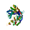 5b5rS S: Starting model for refinement |
|---|---|
| Similar structure data |


-Links
 PDBj
PDBj


- Assembly
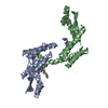
| Deposited unit | 
| ||||||||
|---|---|---|---|---|---|---|---|---|---|
| 1 |
| ||||||||
| Unit cell |
|
-Components
| #1: Protein | Mass: 22511.994 Da / Num. of mol.: 2 / Fragment: C-terminal (UNP RESIDUES 276-484) Source method: isolated from a genetically manipulated source Source: (gene. exp.) Homo sapiens (human) / Gene: GSDMD, DFNA5L, GSDMDC1, FKSG10 / Production host:  Escherichia coli (E. coli) / Strain (production host): BL21(DE3) / References: UniProt: P57764 Escherichia coli (E. coli) / Strain (production host): BL21(DE3) / References: UniProt: P57764#2: Chemical | Glycerol  Mass: 92.094 Da / Num. of mol.: 3 / Source method: obtained synthetically / Formula: C3H8O3 Mass: 92.094 Da / Num. of mol.: 3 / Source method: obtained synthetically / Formula: C3H8O3#3: Chemical | Citric acid  Mass: 192.124 Da / Num. of mol.: 2 / Source method: obtained synthetically / Formula: C6H8O7 Mass: 192.124 Da / Num. of mol.: 2 / Source method: obtained synthetically / Formula: C6H8O7#4: Water | ChemComp-HOH / | Water Mass: 18.015 Da / Num. of mol.: 38 / Source method: isolated from a natural source / Formula: H2O Mass: 18.015 Da / Num. of mol.: 38 / Source method: isolated from a natural source / Formula: H2O |
|---|
-Experimental details
-Experiment
| Experiment | Method: X-RAY DIFFRACTION / Number of used crystals: 1 |
|---|
- Sample preparation
| Crystal | Density Matthews: 2.59 Å3/Da / Density % sol: 52.53 % |
|---|---|
| Crystal grow | Temperature: 291 K / Method: evaporation / pH: 7 Details: 20mM Sodium bromide, 0.1M Hepes sodium pH7.0, 1.5M Sodium citrate tribasic dihydrate |
-Data collection
| Diffraction | Mean temperature: 100 K |
|---|---|
| Diffraction source | Source: SYNCHROTRON / Site: SSRF  / Beamline: BL19U1 / Wavelength: 0.979 Å / Beamline: BL19U1 / Wavelength: 0.979 Å |
| Detector | Type: ADSC QUANTUM 315 / Detector: CCD / Date: Jun 8, 2016 |
| Radiation | Protocol: SINGLE WAVELENGTH / Monochromatic (M) / Laue (L): M / Scattering type: x-ray |
| Radiation wavelength | Wavelength: 0.979 Å / Relative weight: 1 |
| Reflection | Resolution: 2.64→68.33 Å / Num. obs: 13984 / % possible obs: 98 % / Redundancy: 6.4 % / Rmerge(I) obs: 0.194 / Rsym value: 0.194 / Net I/σ(I): 13.25 |
| Reflection shell | Resolution: 2.64→2.7 Å / Redundancy: 6 % / Rmerge(I) obs: 0.694 / Mean I/σ(I) obs: 2.22 / CC1/2: 0.911 / % possible all: 97.6 |
- Processing
| Software |
| ||||||||||||||||||||||||||||||||||||||||||||||||||||||||||||||||||||||||||||||||||||||||||||||||||||||||||||||||||||||||||||||||||||||||||||||||||||||||||||||||||||||||||||||||||||||
|---|---|---|---|---|---|---|---|---|---|---|---|---|---|---|---|---|---|---|---|---|---|---|---|---|---|---|---|---|---|---|---|---|---|---|---|---|---|---|---|---|---|---|---|---|---|---|---|---|---|---|---|---|---|---|---|---|---|---|---|---|---|---|---|---|---|---|---|---|---|---|---|---|---|---|---|---|---|---|---|---|---|---|---|---|---|---|---|---|---|---|---|---|---|---|---|---|---|---|---|---|---|---|---|---|---|---|---|---|---|---|---|---|---|---|---|---|---|---|---|---|---|---|---|---|---|---|---|---|---|---|---|---|---|---|---|---|---|---|---|---|---|---|---|---|---|---|---|---|---|---|---|---|---|---|---|---|---|---|---|---|---|---|---|---|---|---|---|---|---|---|---|---|---|---|---|---|---|---|---|---|---|---|---|
| Refinement | Method to determine structure: MOLECULAR REPLACEMENT Starting model: 5B5R Resolution: 2.64→68.33 Å / Cor.coef. Fo:Fc: 0.942 / Cor.coef. Fo:Fc free: 0.915 / SU B: 14.791 / SU ML: 0.304 / Cross valid method: THROUGHOUT / ESU R: 0.912 / ESU R Free: 0.364 / Details: HYDROGENS HAVE BEEN ADDED IN THE RIDING POSITIONS
| ||||||||||||||||||||||||||||||||||||||||||||||||||||||||||||||||||||||||||||||||||||||||||||||||||||||||||||||||||||||||||||||||||||||||||||||||||||||||||||||||||||||||||||||||||||||
| Solvent computation | Ion probe radii: 0.8 Å / Shrinkage radii: 0.8 Å / VDW probe radii: 1.2 Å | ||||||||||||||||||||||||||||||||||||||||||||||||||||||||||||||||||||||||||||||||||||||||||||||||||||||||||||||||||||||||||||||||||||||||||||||||||||||||||||||||||||||||||||||||||||||
| Displacement parameters | Biso mean: 51.198 Å2
| ||||||||||||||||||||||||||||||||||||||||||||||||||||||||||||||||||||||||||||||||||||||||||||||||||||||||||||||||||||||||||||||||||||||||||||||||||||||||||||||||||||||||||||||||||||||
| Refinement step | Cycle: 1 / Resolution: 2.64→68.33 Å
| ||||||||||||||||||||||||||||||||||||||||||||||||||||||||||||||||||||||||||||||||||||||||||||||||||||||||||||||||||||||||||||||||||||||||||||||||||||||||||||||||||||||||||||||||||||||
| Refine LS restraints |
|