 Movie
Movie Controller


+ Open data

- Basic information
| Entry | Database: PDB / ID: 5wmo | ||||||
|---|---|---|---|---|---|---|---|
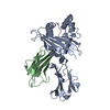
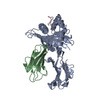
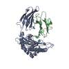
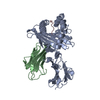
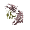
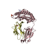
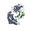
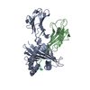
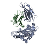
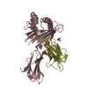
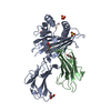
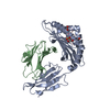
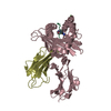
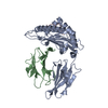
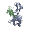
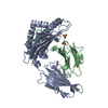
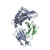
| Title | Crystal Structure of HLA-B7 in complex with RPP, an EBV peptide | ||||||
 Components Components |
| ||||||
 Keywords Keywords |  IMMUNE SYSTEM / HLA / viral peptide / IAV / EBV / CMV / crossreactivity / TCR / T cell / heterologous immunity IMMUNE SYSTEM / HLA / viral peptide / IAV / EBV / CMV / crossreactivity / TCR / T cell / heterologous immunity | ||||||
| Function / homology |  Function and homology information Function and homology informationhost cell nuclear matrix / DNA-templated viral transcription / regulation of interleukin-12 production / regulation of dendritic cell differentiation / regulation of T cell anergy / regulation of interleukin-6 production / TAP binding / protection from natural killer cell mediated cytotoxicity / detection of bacterium / antigen processing and presentation of endogenous peptide antigen via MHC class I via ER pathway, TAP-independent ...host cell nuclear matrix / DNA-templated viral transcription / regulation of interleukin-12 production / regulation of dendritic cell differentiation / regulation of T cell anergy / regulation of interleukin-6 production / TAP binding / protection from natural killer cell mediated cytotoxicity / detection of bacterium / antigen processing and presentation of endogenous peptide antigen via MHC class I via ER pathway, TAP-independent / antigen processing and presentation of endogenous peptide antigen via MHC class Ib / secretory granule membrane / positive regulation of ferrous iron binding / positive regulation of transferrin receptor binding / positive regulation of receptor binding / early endosome lumen / Nef mediated downregulation of MHC class I complex cell surface expression / DAP12 interactions / negative regulation of receptor binding / lumenal side of endoplasmic reticulum membrane / Endosomal/Vacuolar pathway / Antigen Presentation: Folding, assembly and peptide loading of class I MHC / cellular response to iron(III) ion / antigen processing and presentation of exogenous protein antigen via MHC class Ib, TAP-dependent / negative regulation of forebrain neuron differentiation / ER to Golgi transport vesicle membrane / regulation of erythrocyte differentiation / peptide antigen assembly with MHC class I protein complex / response to molecule of bacterial origin / regulation of iron ion transport / MHC class I peptide loading complex / HFE-transferrin receptor complex / T cell mediated cytotoxicity / cellular response to iron ion / antigen processing and presentation of endogenous peptide antigen via MHC class I / positive regulation of T cell cytokine production / defense response / MHC class I protein complex / multicellular organismal-level iron ion homeostasis / negative regulation of neurogenesis / peptide antigen assembly with MHC class II protein complex / positive regulation of receptor-mediated endocytosis / positive regulation of T cell mediated cytotoxicity / MHC class II protein complex / cellular response to nicotine / recycling endosome membrane / specific granule lumen / phagocytic vesicle membrane / peptide antigen binding / positive regulation of cellular senescence / antigen processing and presentation of exogenous peptide antigen via MHC class II / negative regulation of epithelial cell proliferation / Immunoregulatory interactions between a Lymphoid and a non-Lymphoid cell / Interferon gamma signaling / positive regulation of immune response / Modulation by Mtb of host immune system / Interferon alpha/beta signaling / sensory perception of smell / positive regulation of T cell activation / positive regulation of protein binding / tertiary granule lumen / DAP12 signaling / negative regulation of neuron projection development / MHC class II protein complex binding / late endosome membrane / T cell differentiation in thymus / ER-Phagosome pathway / iron ion transport / protein-folding chaperone binding / early endosome membrane / protein refolding / protein homotetramerization / intracellular iron ion homeostasis / adaptive immune response / amyloid fibril formation / learning or memory / immune response / Amyloid fiber formation / lysosomal membrane / external side of plasma membrane / endoplasmic reticulum lumen / Golgi membrane / signaling receptor binding / focal adhesion / innate immune response / Neutrophil degranulation / SARS-CoV-2 activates/modulates innate and adaptive immune responses / structural molecule activity / Golgi apparatus / cell surface / endoplasmic reticulum / protein homodimerization activity / extracellular space / extracellular exosome / extracellular region / membrane / identical protein binding / plasma membrane / cytosolSimilarity search - Function | ||||||
| Biological species |  Homo sapiens (human) Homo sapiens (human) Epstein-barr virus strain ag876 Epstein-barr virus strain ag876 | ||||||
| Method | X-RAY DIFFRACTION / SYNCHROTRON / MOLECULAR REPLACEMENT / molecular replacement / Resolution: 1.62 Å | ||||||
 Authors Authors | Gras, S. / Rossjohn, J. | ||||||
 Citation Citation | Journal: J. Immunol. / Year: 2018 Title: Inability To Detect Cross-Reactive Memory T Cells Challenges the Frequency of Heterologous Immunity among Common Viruses. Authors: Rowntree, L.C. / Nguyen, T.H.O. / Halim, H. / Purcell, A.W. / Rossjohn, J. / Gras, S. / Kotsimbos, T.C. / Mifsud, N.A. | ||||||
| History |
|
- Structure visualization
| Structure viewer | Molecule: MolmilJmol/JSmol |
|---|
- Downloads & links
-Download
| PDBx/mmCIF format | 5wmo.cif.gz | 113.3 KB | Display | PDBx/mmCIF format |
|---|---|---|---|---|
| PDB format | pdb5wmo.ent.gz | 84 KB | Display | PDB format |
| PDBx/mmJSON format | 5wmo.json.gz | Tree view | PDBx/mmJSON format | |
| Others |  Other downloads Other downloads |
-Validation report
| Arichive directory | https://data.pdbj.org/pub/pdb/validation_reports/wm/5wmoftp://data.pdbj.org/pub/pdb/validation_reports/wm/5wmo | HTTPS FTP |
|---|
-Related structure data
| Related structure data | 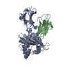 5wmnC 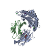 5wmpC  5wmqC  5wmrC  3vclS S: Starting model for refinement C: citing same article ( |
|---|---|
| Similar structure data |


-Links
 PDBj
PDBj

- Assembly
| Deposited unit | 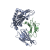
| ||||||||
|---|---|---|---|---|---|---|---|---|---|
| 1 |
| ||||||||
| Unit cell |
|
-Components
| #1: Protein | Mass: 32059.129 Da / Num. of mol.: 1 Source method: isolated from a genetically manipulated source Source: (gene. exp.) Homo sapiens (human) / Gene: HLA-B, HLAB / Plasmid: pET30 / Production host:  Escherichia coli (E. coli) / Strain (production host): BL21(DE3) / References: UniProt: P01889 Escherichia coli (E. coli) / Strain (production host): BL21(DE3) / References: UniProt: P01889 |
|---|---|
| #2: Protein | Beta-2 microglobulin Mass: 11820.270 Da / Num. of mol.: 1 Source method: isolated from a genetically manipulated source Source: (gene. exp.) Homo sapiens (human) / Gene: B2M, CDABP0092, HDCMA22P / Plasmid: pET30 / Production host: Escherichia coli (E. coli) / Strain (production host): BL21(DE3) / References: UniProt: P61769 |
| #3: Protein/peptide | Mass: 1170.473 Da / Num. of mol.: 1 / Source method: obtained synthetically / Source: (synth.) Epstein-barr virus strain ag876 / References: UniProt: P12977*PLUS |
| #4: Water | ChemComp-HOH / Water Mass: 18.015 Da / Num. of mol.: 658 / Source method: isolated from a natural source / Formula: H2O Mass: 18.015 Da / Num. of mol.: 658 / Source method: isolated from a natural source / Formula: H2O |
-Experimental details
-Experiment
| Experiment | Method: X-RAY DIFFRACTION / Number of used crystals: 1 |
|---|
- Sample preparation
| Crystal | Density Matthews: 2.52 Å3/Da / Density % sol: 51.1 % / Mosaicity: 0.09 ° |
|---|---|
| Crystal grow | Temperature: 277 K / Method: vapor diffusion, hanging drop / pH: 6.5 Details: 18-24%PEG4000, 0.2 NH4 Acetate, 0.1M Na-cacodylate pH 6.5 |
-Data collection
| Diffraction | Mean temperature: 100 K | ||||||||||||||||||||||||||||||
|---|---|---|---|---|---|---|---|---|---|---|---|---|---|---|---|---|---|---|---|---|---|---|---|---|---|---|---|---|---|---|---|
| Diffraction source | Source: SYNCHROTRON / Site: Australian Synchrotron  / Beamline: MX2 / Wavelength: 0.954 Å / Beamline: MX2 / Wavelength: 0.954 Å | ||||||||||||||||||||||||||||||
| Detector | Type: ADSC QUANTUM 315r / Detector: CCD / Date: Feb 8, 2014 | ||||||||||||||||||||||||||||||
| Radiation | Protocol: SINGLE WAVELENGTH / Monochromatic (M) / Laue (L): M / Scattering type: x-ray | ||||||||||||||||||||||||||||||
| Radiation wavelength | Wavelength: 0.954 Å / Relative weight: 1 | ||||||||||||||||||||||||||||||
| Reflection | Resolution: 1.62→45.92 Å / Num. all: 58022 / Num. obs: 58022 / % possible obs: 99.8 % / Redundancy: 7.3 % / Biso Wilson estimate: 17.65 Å2 / Rmerge(I) obs: 0.108 / Rpim(I) all: 0.043 / Rrim(I) all: 0.116 / Rsym value: 0.108 / Net I/σ(I): 15.1 / Num. measured all: 423294 | ||||||||||||||||||||||||||||||
| Reflection shell | Diffraction-ID: 1
|
-Phasing
| Phasing | Method: molecular replacement |
|---|
- Processing
| Software |
| ||||||||||||||||||||||||||||||||||||||||||||||||||||||||||||||||||||||||||||||||||||||||||||||||||||||||||||
|---|---|---|---|---|---|---|---|---|---|---|---|---|---|---|---|---|---|---|---|---|---|---|---|---|---|---|---|---|---|---|---|---|---|---|---|---|---|---|---|---|---|---|---|---|---|---|---|---|---|---|---|---|---|---|---|---|---|---|---|---|---|---|---|---|---|---|---|---|---|---|---|---|---|---|---|---|---|---|---|---|---|---|---|---|---|---|---|---|---|---|---|---|---|---|---|---|---|---|---|---|---|---|---|---|---|---|---|---|---|
| Refinement | Method to determine structure: MOLECULAR REPLACEMENT Starting model: 3vcl Resolution: 1.62→45.92 Å / Cor.coef. Fo:Fc: 0.9534 / Cor.coef. Fo:Fc free: 0.9406 / SU R Cruickshank DPI: 0.086 / Cross valid method: THROUGHOUT / σ(F): 0 / SU R Blow DPI: 0.095 / SU Rfree Blow DPI: 0.091 / SU Rfree Cruickshank DPI: 0.085
| ||||||||||||||||||||||||||||||||||||||||||||||||||||||||||||||||||||||||||||||||||||||||||||||||||||||||||||
| Displacement parameters | Biso max: 88.11 Å2 / Biso mean: 20.27 Å2 / Biso min: 4.54 Å2
| ||||||||||||||||||||||||||||||||||||||||||||||||||||||||||||||||||||||||||||||||||||||||||||||||||||||||||||
| Refine analyze | Luzzati coordinate error obs: 0.165 Å | ||||||||||||||||||||||||||||||||||||||||||||||||||||||||||||||||||||||||||||||||||||||||||||||||||||||||||||
| Refinement step | Cycle: final / Resolution: 1.62→45.92 Å
| ||||||||||||||||||||||||||||||||||||||||||||||||||||||||||||||||||||||||||||||||||||||||||||||||||||||||||||
| Refine LS restraints |
| ||||||||||||||||||||||||||||||||||||||||||||||||||||||||||||||||||||||||||||||||||||||||||||||||||||||||||||
| LS refinement shell | Resolution: 1.62→1.66 Å / Rfactor Rfree error: 0 / Total num. of bins used: 20
|
