 Movie
Movie Controller

[English] 日本語
 Yorodumi
- PDB-4u1k: HLA class I micropolymorphisms determine peptide-HLA landscape an... -
+ Open data

- Basic information
| Entry | Database: PDB / ID: 4u1k | |||||||||
|---|---|---|---|---|---|---|---|---|---|---|
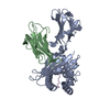
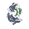
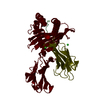
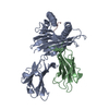
| Title | HLA class I micropolymorphisms determine peptide-HLA landscape and dictate differential HIV-1 escape through identical epitopes | |||||||||
 Components Components |
| |||||||||
 Keywords Keywords |  IMMUNE SYSTEM / Immunoglobulin / HLA / HIV IMMUNE SYSTEM / Immunoglobulin / HLA / HIV | |||||||||
| Function / homology |  Function and homology information Function and homology informationsymbiont-mediated suppression of host antigen processing and presentation of peptide antigen via MHC class I / symbiont-mediated suppression of host antigen processing and presentation of peptide antigen via MHC class II / suppression by virus of host autophagy / regulation of interleukin-12 production / regulation of dendritic cell differentiation / regulation of T cell anergy / activation of transmembrane receptor protein tyrosine kinase activity / regulation of interleukin-6 production / host cell Golgi membrane / host cell membrane ...symbiont-mediated suppression of host antigen processing and presentation of peptide antigen via MHC class I / symbiont-mediated suppression of host antigen processing and presentation of peptide antigen via MHC class II / suppression by virus of host autophagy / regulation of interleukin-12 production / regulation of dendritic cell differentiation / regulation of T cell anergy / activation of transmembrane receptor protein tyrosine kinase activity / regulation of interleukin-6 production / host cell Golgi membrane / host cell membrane / TAP binding / protection from natural killer cell mediated cytotoxicity / antigen processing and presentation of endogenous peptide antigen via MHC class I via ER pathway, TAP-independent / antigen processing and presentation of endogenous peptide antigen via MHC class Ib / detection of bacterium / secretory granule membrane / virus-mediated perturbation of host defense response / positive regulation of ferrous iron binding / positive regulation of transferrin receptor binding / positive regulation of receptor binding / early endosome lumen / Nef mediated downregulation of MHC class I complex cell surface expression / DAP12 interactions / negative regulation of receptor binding / virion component / lumenal side of endoplasmic reticulum membrane / Endosomal/Vacuolar pathway / Antigen Presentation: Folding, assembly and peptide loading of class I MHC / antigen processing and presentation of exogenous protein antigen via MHC class Ib, TAP-dependent / cellular response to iron(III) ion / negative regulation of forebrain neuron differentiation / ER to Golgi transport vesicle membrane / peptide antigen assembly with MHC class I protein complex / response to molecule of bacterial origin / regulation of erythrocyte differentiation / endocytosis involved in viral entry into host cell / regulation of iron ion transport / MHC class I peptide loading complex / HFE-transferrin receptor complex / T cell mediated cytotoxicity / cellular response to iron ion / antigen processing and presentation of endogenous peptide antigen via MHC class I / positive regulation of T cell cytokine production / defense response / MHC class I protein complex / multicellular organismal-level iron ion homeostasis / SH3 domain binding / positive regulation of T cell mediated cytotoxicity / peptide antigen assembly with MHC class II protein complex / negative regulation of neurogenesis / MHC class II protein complex / positive regulation of receptor-mediated endocytosis / cellular response to nicotine / recycling endosome membrane / specific granule lumen / phagocytic vesicle membrane / peptide antigen binding / positive regulation of cellular senescence / antigen processing and presentation of exogenous peptide antigen via MHC class II / negative regulation of epithelial cell proliferation / Immunoregulatory interactions between a Lymphoid and a non-Lymphoid cell / positive regulation of immune response / Interferon gamma signaling / Modulation by Mtb of host immune system / positive regulation of T cell activation / Interferon alpha/beta signaling / sensory perception of smell / negative regulation of neuron projection development / tertiary granule lumen / DAP12 signaling / positive regulation of protein binding / MHC class II protein complex binding / T cell differentiation in thymus / late endosome membrane / ER-Phagosome pathway / iron ion transport / protein-folding chaperone binding / protein refolding / early endosome membrane / protein homotetramerization / intracellular iron ion homeostasis / amyloid fibril formation / adaptive immune response / learning or memory / immune response / Amyloid fiber formation / lysosomal membrane / external side of plasma membrane / endoplasmic reticulum lumen / Golgi membrane / signaling receptor binding / focal adhesion / innate immune response / Neutrophil degranulation / GTP binding / SARS-CoV-2 activates/modulates innate and adaptive immune responses / host cell plasma membrane / structural molecule activity / Golgi apparatus / cell surfaceSimilarity search - Function | |||||||||
| Biological species |  Homo sapiens (human) Homo sapiens (human)  Human immunodeficiency virus 1 Human immunodeficiency virus 1 | |||||||||
| Method | X-RAY DIFFRACTION / SYNCHROTRON / MOLECULAR REPLACEMENT / molecular replacement / Resolution: 2.09 Å | |||||||||
| Model details | HLA-B0702 carrying RM9 peptide | |||||||||
 Authors Authors | Rizkallah, P.J. / Cole, D.K. / Fuller, A. / Sewell, A.K. | |||||||||
| Funding support |  United Kingdom, 1items United Kingdom, 1items
| |||||||||
 Citation Citation | Journal: Retrovirology / Year: 2015 Title: A molecular switch in immunodominant HIV-1-specific CD8 T-cell epitopes shapes differential HLA-restricted escape. Authors: Klverpris, H.N. / Cole, D.K. / Fuller, A. / Carlson, J. / Beck, K. / Schauenburg, A.J. / Rizkallah, P.J. / Buus, S. / Sewell, A.K. / Goulder, P. | |||||||||
| History |
|
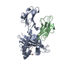
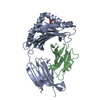
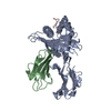
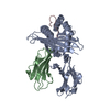
- Structure visualization
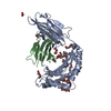
| Structure viewer | Molecule: MolmilJmol/JSmol |
|---|
- Downloads & links
-Download
| PDBx/mmCIF format | 4u1k.cif.gz | 348.7 KB | Display | PDBx/mmCIF format |
|---|---|---|---|---|
| PDB format | pdb4u1k.ent.gz | 284.1 KB | Display | PDB format |
| PDBx/mmJSON format | 4u1k.json.gz | Tree view | PDBx/mmJSON format | |
| Others |  Other downloads Other downloads |
-Validation report
| Arichive directory | https://data.pdbj.org/pub/pdb/validation_reports/u1/4u1kftp://data.pdbj.org/pub/pdb/validation_reports/u1/4u1k | HTTPS FTP |
|---|
-Related structure data
| Related structure data |  4u1hC  4u1iC  4u1jC  4u1lC  4u1mC  4u1nC  4u1sC  4i4wS C: citing same article ( S: Starting model for refinement |
|---|---|
| Similar structure data |


-Links
 PDBj
PDBj


- Assembly
| Deposited unit | 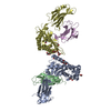
| ||||||||
|---|---|---|---|---|---|---|---|---|---|
| 1 | 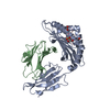
| ||||||||
| 2 | 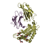
| ||||||||
| Unit cell |
| ||||||||
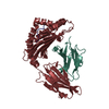
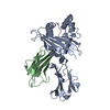
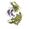
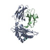
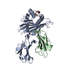
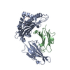
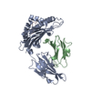
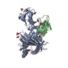
| Details | Chains A, B and C form one biological entity. A second copy in the a.u. is formed by chains D, E and F |
-Components
-Protein , 2 types, 4 molecules ADBE
| #1: Protein | Mass: 32190.324 Da / Num. of mol.: 2 / Fragment: UNP residues 25-300 Source method: isolated from a genetically manipulated source Source: (gene. exp.) Homo sapiens (human) / Gene: HLA-B, HLAB / Plasmid: pGMT7 / Production host:  Escherichia coli BL21(DE3) (bacteria) / Strain (production host): Rosetta / References: UniProt: P01889 Escherichia coli BL21(DE3) (bacteria) / Strain (production host): Rosetta / References: UniProt: P01889#2: Protein | Beta-2 microglobulinMass: 11879.356 Da / Num. of mol.: 2 / Fragment: UNP residues 21-119 Source method: isolated from a genetically manipulated source Source: (gene. exp.) Homo sapiens (human) / Gene: B2M, CDABP0092, HDCMA22P / Plasmid: pGMT7 / Production host: Escherichia coli BL21(DE3) (bacteria) / Strain (production host): Rosetta / References: UniProt: P61769 |
|---|
-Protein/peptide , 1 types, 2 molecules CF
| #3: Protein/peptide | Mass: 1095.360 Da / Num. of mol.: 2 / Source method: obtained synthetically / Source: (synth.) Human immunodeficiency virus 1 / References: UniProt: Q90VG9, UniProt: P03407*PLUS |
|---|
-Non-polymers , 4 types, 512 molecules 


| #4: Chemical | ChemComp-GOL / Glycerol Mass: 92.094 Da / Num. of mol.: 8 / Source method: obtained synthetically / Formula: C3H8O3 Mass: 92.094 Da / Num. of mol.: 8 / Source method: obtained synthetically / Formula: C3H8O3#5: Chemical | ChemComp-EDO / Ethylene glycol Mass: 62.068 Da / Num. of mol.: 6 / Source method: obtained synthetically / Formula: C2H6O2 Mass: 62.068 Da / Num. of mol.: 6 / Source method: obtained synthetically / Formula: C2H6O2#6: Chemical | ChemComp-SO4 / | Sulfate Mass: 96.063 Da / Num. of mol.: 1 / Source method: obtained synthetically / Formula: SO4 Mass: 96.063 Da / Num. of mol.: 1 / Source method: obtained synthetically / Formula: SO4#7: Water | ChemComp-HOH / | WaterMass: 18.015 Da / Num. of mol.: 497 / Source method: isolated from a natural source / Formula: H2O |
|---|
-Experimental details
-Experiment
| Experiment | Method: X-RAY DIFFRACTION / Number of used crystals: 1 |
|---|
- Sample preparation
| Crystal | Density Matthews: 2.68 Å3/Da / Density % sol: 54.18 % |
|---|---|
| Crystal grow | Temperature: 291 K / Method: vapor diffusion, sitting drop / pH: 6 Details: 0.1 M Sodium Cacodylate, pH 6, 15% PEG 8000, 15% Glycerol |
-Data collection
| Diffraction | Mean temperature: 100 K | |||||||||||||||||||||||||||
|---|---|---|---|---|---|---|---|---|---|---|---|---|---|---|---|---|---|---|---|---|---|---|---|---|---|---|---|---|
| Diffraction source | Source: SYNCHROTRON / Site: Diamond / Beamline: I03 / Wavelength: 0.9763 Å | |||||||||||||||||||||||||||
| Detector | Type: PSI PILATUS 6M / Detector: PIXEL / Date: Jun 30, 2012 / Details: mirrors | |||||||||||||||||||||||||||
| Radiation | Protocol: SINGLE WAVELENGTH / Monochromatic (M) / Laue (L): M / Scattering type: x-ray | |||||||||||||||||||||||||||
| Radiation wavelength | Wavelength: 0.9763 Å / Relative weight: 1 | |||||||||||||||||||||||||||
| Reflection | Resolution: 2.09→48.86 Å / Num. obs: 54249 / % possible obs: 97.5 % / Redundancy: 2 % / CC1/2: 0.983 / Rmerge(I) obs: 0.098 / Rpim(I) all: 0.087 / Net I/σ(I): 4.8 / Num. measured all: 107642 | |||||||||||||||||||||||||||
| Reflection shell | Diffraction-ID: 1 / Rejects: 0
|
-Phasing
| Phasing | Method: molecular replacement | |||||||||
|---|---|---|---|---|---|---|---|---|---|---|
| Phasing MR | Model details: Phaser MODE: MR_AUTO
|
- Processing
| Software |
| |||||||||||||||||||||||||||||||||||||||||||||||||||||||||||||||||||||||||||||||||||||||||||||||||||||||||||||||||||||||||||||||||||||||||||||||||||||||||||||||||||||||||||||||
|---|---|---|---|---|---|---|---|---|---|---|---|---|---|---|---|---|---|---|---|---|---|---|---|---|---|---|---|---|---|---|---|---|---|---|---|---|---|---|---|---|---|---|---|---|---|---|---|---|---|---|---|---|---|---|---|---|---|---|---|---|---|---|---|---|---|---|---|---|---|---|---|---|---|---|---|---|---|---|---|---|---|---|---|---|---|---|---|---|---|---|---|---|---|---|---|---|---|---|---|---|---|---|---|---|---|---|---|---|---|---|---|---|---|---|---|---|---|---|---|---|---|---|---|---|---|---|---|---|---|---|---|---|---|---|---|---|---|---|---|---|---|---|---|---|---|---|---|---|---|---|---|---|---|---|---|---|---|---|---|---|---|---|---|---|---|---|---|---|---|---|---|---|---|---|---|---|
| Refinement | Method to determine structure: MOLECULAR REPLACEMENT Starting model: 4I4W Resolution: 2.09→48.86 Å / Cor.coef. Fo:Fc: 0.953 / Cor.coef. Fo:Fc free: 0.919 / WRfactor Rfree: 0.2393 / WRfactor Rwork: 0.185 / FOM work R set: 0.8298 / SU B: 10.564 / SU ML: 0.145 / SU R Cruickshank DPI: 0.2055 / SU Rfree: 0.1789 / Cross valid method: THROUGHOUT / σ(F): 0 / ESU R: 0.205 / ESU R Free: 0.179 / Stereochemistry target values: MAXIMUM LIKELIHOOD Details: HYDROGENS HAVE BEEN ADDED IN THE RIDING POSITIONS U VALUES : WITH TLS ADDED
| |||||||||||||||||||||||||||||||||||||||||||||||||||||||||||||||||||||||||||||||||||||||||||||||||||||||||||||||||||||||||||||||||||||||||||||||||||||||||||||||||||||||||||||||
| Solvent computation | Ion probe radii: 0.8 Å / Shrinkage radii: 0.8 Å / VDW probe radii: 1.2 Å / Solvent model: MASK | |||||||||||||||||||||||||||||||||||||||||||||||||||||||||||||||||||||||||||||||||||||||||||||||||||||||||||||||||||||||||||||||||||||||||||||||||||||||||||||||||||||||||||||||
| Displacement parameters | Biso max: 148.82 Å2 / Biso mean: 51.539 Å2 / Biso min: 14.57 Å2
| |||||||||||||||||||||||||||||||||||||||||||||||||||||||||||||||||||||||||||||||||||||||||||||||||||||||||||||||||||||||||||||||||||||||||||||||||||||||||||||||||||||||||||||||
| Refinement step | Cycle: final / Resolution: 2.09→48.86 Å
| |||||||||||||||||||||||||||||||||||||||||||||||||||||||||||||||||||||||||||||||||||||||||||||||||||||||||||||||||||||||||||||||||||||||||||||||||||||||||||||||||||||||||||||||
| Refine LS restraints |
| |||||||||||||||||||||||||||||||||||||||||||||||||||||||||||||||||||||||||||||||||||||||||||||||||||||||||||||||||||||||||||||||||||||||||||||||||||||||||||||||||||||||||||||||
| LS refinement shell | Resolution: 2.09→2.144 Å / Total num. of bins used: 20
| |||||||||||||||||||||||||||||||||||||||||||||||||||||||||||||||||||||||||||||||||||||||||||||||||||||||||||||||||||||||||||||||||||||||||||||||||||||||||||||||||||||||||||||||
| Refinement TLS params. | Method: refined / Refine-ID: X-RAY DIFFRACTION
| |||||||||||||||||||||||||||||||||||||||||||||||||||||||||||||||||||||||||||||||||||||||||||||||||||||||||||||||||||||||||||||||||||||||||||||||||||||||||||||||||||||||||||||||
| Refinement TLS group |
|
