 Movie
Movie Controller

[English] 日本語
 Yorodumi
- PDB-5wka: Crystal structure of a GH1 beta-glucosidase retrieved from microb... -
+ Open data

- Basic information
| Entry | Database: PDB / ID: 5wka | ||||||
|---|---|---|---|---|---|---|---|
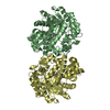
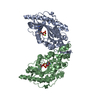
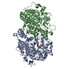
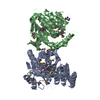
| Title | Crystal structure of a GH1 beta-glucosidase retrieved from microbial metagenome of Poraque Amazon lake | ||||||
 Components Components | Beta-glucosidase | ||||||
 Keywords Keywords | HYDROLASE / Beta-glucosidase / GH1-family / metagenome | ||||||
| Function / homology | Glycosidases / TIM Barrel / Alpha-Beta Barrel / Alpha Beta / DI(HYDROXYETHYL)ETHER Function and homology information Function and homology information | ||||||
| Biological species | metagenome (others) | ||||||
| Method | X-RAY DIFFRACTION / SYNCHROTRON / MOLECULAR REPLACEMENT / Resolution: 2.75 Å | ||||||
 Authors Authors | Morais, M.A.B. / Toyama, D. / Ramos, F.C. / Zanphorlin, L.M. / Tonoli, C.C.C. / Miranda, F.P. / Ruller, R. / Henrique-Silva, F. / Murakami, M.T. | ||||||
| Funding support |  Brazil, 1items Brazil, 1items
| ||||||
 Citation Citation | Journal: Biochim. Biophys. Acta / Year: 2018 Title: A novel beta-glucosidase isolated from the microbial metagenome of Lake Poraque (Amazon, Brazil). Authors: Toyama, D. / de Morais, M.A.B. / Ramos, F.C. / Zanphorlin, L.M. / Tonoli, C.C.C. / Balula, A.F. / de Miranda, F.P. / Almeida, V.M. / Marana, S.R. / Ruller, R. / Murakami, M.T. / Henrique-Silva, F. | ||||||
| History |
|
- Structure visualization
| Structure viewer | Molecule: MolmilJmol/JSmol |
|---|
- Downloads & links
-Download
| PDBx/mmCIF format | 5wka.cif.gz | 362.7 KB | Display | PDBx/mmCIF format |
|---|---|---|---|---|
| PDB format | pdb5wka.ent.gz | 297.7 KB | Display | PDB format |
| PDBx/mmJSON format | 5wka.json.gz | Tree view | PDBx/mmJSON format | |
| Others |  Other downloads Other downloads |
-Validation report
| Arichive directory | https://data.pdbj.org/pub/pdb/validation_reports/wk/5wkaftp://data.pdbj.org/pub/pdb/validation_reports/wk/5wka | HTTPS FTP |
|---|
-Related structure data
| Related structure data |  1gnxS S: Starting model for refinement |
|---|---|
| Similar structure data |


-Links
 PDBj
PDBj- Assembly
| Deposited unit | 
| ||||||||
|---|---|---|---|---|---|---|---|---|---|
| 1 | 
| ||||||||
| 2 | 
| ||||||||
| Unit cell |
| ||||||||
| Details | homodimer by SAXS |
-Components
| #1: Protein | Mass: 52977.625 Da / Num. of mol.: 4 Source method: isolated from a genetically manipulated source Source: (gene. exp.) metagenome (others) / Production host:  Escherichia coli (E. coli) Escherichia coli (E. coli)#2: Chemical | ChemComp-GOL / Glycerol  Mass: 92.094 Da / Num. of mol.: 6 / Source method: obtained synthetically / Formula: C3H8O3 Mass: 92.094 Da / Num. of mol.: 6 / Source method: obtained synthetically / Formula: C3H8O3#3: Chemical | ChemComp-PEG / Diethylene glycol  Mass: 106.120 Da / Num. of mol.: 4 / Source method: obtained synthetically / Formula: C4H10O3 Mass: 106.120 Da / Num. of mol.: 4 / Source method: obtained synthetically / Formula: C4H10O3#4: Water | ChemComp-HOH / | Water Mass: 18.015 Da / Num. of mol.: 10 / Source method: isolated from a natural source / Formula: H2O Mass: 18.015 Da / Num. of mol.: 10 / Source method: isolated from a natural source / Formula: H2O |
|---|
-Experimental details
-Experiment
| Experiment | Method: X-RAY DIFFRACTION / Number of used crystals: 1 |
|---|
- Sample preparation
| Crystal | Density Matthews: 2.3 Å3/Da / Density % sol: 46.56 % |
|---|---|
| Crystal grow | Temperature: 291 K / Method: vapor diffusion, sitting drop Details: 0.1 M MES pH 6.0, PEG 6000 20% (w/v) and 0.2 M sodium chloride. |
-Data collection
| Diffraction | Mean temperature: 100 K | ||||||||||||
|---|---|---|---|---|---|---|---|---|---|---|---|---|---|
| Diffraction source | Source: SYNCHROTRON / Site: LNLS / Beamline: W01B-MX2 / Wavelength: 1.459 Å | ||||||||||||
| Detector | Type: DECTRIS PILATUS 2M / Detector: PIXEL / Date: Sep 27, 2016 | ||||||||||||
| Radiation | Protocol: SINGLE WAVELENGTH / Monochromatic (M) / Laue (L): M / Scattering type: x-ray | ||||||||||||
| Radiation wavelength | Wavelength: 1.459 Å / Relative weight: 1 | ||||||||||||
| Reflection | Resolution: 2.75→46.463 Å / Num. obs: 49452 / % possible obs: 88.17 % / Redundancy: 3 % / Biso Wilson estimate: 57 Å2 / Net I/σ(I): 6 | ||||||||||||
| Reflection shell |
|
- Processing
| Software |
| ||||||||||||||||||||||||||||||||||||||||||||||||||||||||||||||||||||||||||||||||||||||||||||||||||||||||||||||||||||||||||||||||||||||||||||||||||||||||||||||||||||||||||||||||||||||||||||||||||||||||||||||||||
|---|---|---|---|---|---|---|---|---|---|---|---|---|---|---|---|---|---|---|---|---|---|---|---|---|---|---|---|---|---|---|---|---|---|---|---|---|---|---|---|---|---|---|---|---|---|---|---|---|---|---|---|---|---|---|---|---|---|---|---|---|---|---|---|---|---|---|---|---|---|---|---|---|---|---|---|---|---|---|---|---|---|---|---|---|---|---|---|---|---|---|---|---|---|---|---|---|---|---|---|---|---|---|---|---|---|---|---|---|---|---|---|---|---|---|---|---|---|---|---|---|---|---|---|---|---|---|---|---|---|---|---|---|---|---|---|---|---|---|---|---|---|---|---|---|---|---|---|---|---|---|---|---|---|---|---|---|---|---|---|---|---|---|---|---|---|---|---|---|---|---|---|---|---|---|---|---|---|---|---|---|---|---|---|---|---|---|---|---|---|---|---|---|---|---|---|---|---|---|---|---|---|---|---|---|---|---|---|---|---|---|---|
| Refinement | Method to determine structure: MOLECULAR REPLACEMENT Starting model: 1GNX Resolution: 2.75→46.463 Å / SU ML: 0.48 / Cross valid method: FREE R-VALUE / σ(F): 1.21 / Phase error: 32.55
| ||||||||||||||||||||||||||||||||||||||||||||||||||||||||||||||||||||||||||||||||||||||||||||||||||||||||||||||||||||||||||||||||||||||||||||||||||||||||||||||||||||||||||||||||||||||||||||||||||||||||||||||||||
| Solvent computation | Shrinkage radii: 0.9 Å / VDW probe radii: 1.11 Å | ||||||||||||||||||||||||||||||||||||||||||||||||||||||||||||||||||||||||||||||||||||||||||||||||||||||||||||||||||||||||||||||||||||||||||||||||||||||||||||||||||||||||||||||||||||||||||||||||||||||||||||||||||
| Displacement parameters | Biso max: 96.83 Å2 / Biso mean: 56.983 Å2 / Biso min: 36.9 Å2 | ||||||||||||||||||||||||||||||||||||||||||||||||||||||||||||||||||||||||||||||||||||||||||||||||||||||||||||||||||||||||||||||||||||||||||||||||||||||||||||||||||||||||||||||||||||||||||||||||||||||||||||||||||
| Refinement step | Cycle: final / Resolution: 2.75→46.463 Å
| ||||||||||||||||||||||||||||||||||||||||||||||||||||||||||||||||||||||||||||||||||||||||||||||||||||||||||||||||||||||||||||||||||||||||||||||||||||||||||||||||||||||||||||||||||||||||||||||||||||||||||||||||||
| Refine LS restraints |
| ||||||||||||||||||||||||||||||||||||||||||||||||||||||||||||||||||||||||||||||||||||||||||||||||||||||||||||||||||||||||||||||||||||||||||||||||||||||||||||||||||||||||||||||||||||||||||||||||||||||||||||||||||
| LS refinement shell | Refine-ID: X-RAY DIFFRACTION / Rfactor Rfree error: 0 / Total num. of bins used: 29
|
