 Movie
Movie Controller


+ Open data

- Basic information
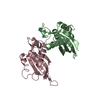
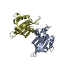
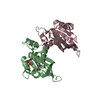
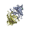
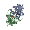
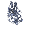
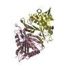
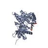
| Entry | Database: PDB / ID: 5wip | ||||||
|---|---|---|---|---|---|---|---|
| Title | TraE protein in complex with 2-(2-furyl)isonicotinic acid | ||||||
 Components Components | Conjugal transfer protein | ||||||
 Keywords Keywords |  PROTEIN TRANSPORT / Type IV Secretion System / Fragment-Based Drug Design / Complex / Inhibitor / Antimicrobial Resistance / Bacterial secretion / VirB PROTEIN TRANSPORT / Type IV Secretion System / Fragment-Based Drug Design / Complex / Inhibitor / Antimicrobial Resistance / Bacterial secretion / VirB | ||||||
| Function / homology |  Function and homology information Function and homology information | ||||||
| Biological species |  Escherichia coli (E. coli) Escherichia coli (E. coli) | ||||||
| Method | X-RAY DIFFRACTION / SYNCHROTRON / MOLECULAR REPLACEMENT / Resolution: 2.62 Å | ||||||
 Authors Authors | Casu, B. / Arya, T. / Bessette, B. / Baron, C. | ||||||
| Funding support |  Canada, 1items Canada, 1items
| ||||||
 Citation Citation | Journal: Sci Rep / Year: 2017 Title: Fragment-based screening identifies novel targets for inhibitors of conjugative transfer of antimicrobial resistance by plasmid pKM101. Authors: Casu, B. / Arya, T. / Bessette, B. / Baron, C. | ||||||
| History |
|
- Structure visualization
| Structure viewer | Molecule: MolmilJmol/JSmol |
|---|
- Downloads & links
-Download
| PDBx/mmCIF format | 5wip.cif.gz | 126.8 KB | Display | PDBx/mmCIF format |
|---|---|---|---|---|
| PDB format | pdb5wip.ent.gz | 98.3 KB | Display | PDB format |
| PDBx/mmJSON format | 5wip.json.gz | Tree view | PDBx/mmJSON format | |
| Others |  Other downloads Other downloads |
-Validation report
| Arichive directory | https://data.pdbj.org/pub/pdb/validation_reports/wi/5wipftp://data.pdbj.org/pub/pdb/validation_reports/wi/5wip | HTTPS FTP |
|---|
-Related structure data
| Related structure data |  5wicC  5wiiC  5wioC  5i97S S: Starting model for refinement C: citing same article ( |
|---|---|
| Similar structure data |

-Links
 PDBj
PDBj

- Assembly
| Deposited unit | 
| ||||||||
|---|---|---|---|---|---|---|---|---|---|
| 1 | 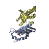
| ||||||||
| 2 | 
| ||||||||
| Unit cell |
|
-Components
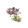
| #1: Protein | Mass: 18818.104 Da / Num. of mol.: 4 / Fragment: residues 70-232 Source method: isolated from a genetically manipulated source Source: (gene. exp.) Escherichia coli (E. coli)Gene: traE, B634_00049, BE957_00530, ECONIH1_27615, pec386IL_00125, pEcNDM0_00049, pHKU1_33, pKC394-028, pKC396-021, pMUR050_041, pN3_023 Plasmid: PHT / Cell line (production host): BL21(DE3)STAR / Production host: Escherichia coli (E. coli) / References: UniProt: Q17U16#2: Chemical | ChemComp-XXO / |   Mass: 189.167 Da / Num. of mol.: 1 / Source method: obtained synthetically / Formula: C10H7NO3 Mass: 189.167 Da / Num. of mol.: 1 / Source method: obtained synthetically / Formula: C10H7NO3#3: Water | ChemComp-HOH / | Water Mass: 18.015 Da / Num. of mol.: 146 / Source method: isolated from a natural source / Formula: H2O Mass: 18.015 Da / Num. of mol.: 146 / Source method: isolated from a natural source / Formula: H2O |
|---|
-Experimental details
-Experiment
| Experiment | Method: X-RAY DIFFRACTION / Number of used crystals: 1 |
|---|
- Sample preparation
| Crystal | Density Matthews: 2.55 Å3/Da / Density % sol: 51.68 % |
|---|---|
| Crystal grow | Temperature: 293 K / Method: vapor diffusion, hanging drop / pH: 7.4 Details: 16% (w/v) PEG 10,000, 50 mM Bis-Tris (pH 5.5), 100 mM ammonium acetate |
-Data collection
| Diffraction | Mean temperature: 100 K |
|---|---|
| Diffraction source | Source: SYNCHROTRON / Site: CHESS  / Beamline: F1 / Wavelength: 0.977 Å / Beamline: F1 / Wavelength: 0.977 Å |
| Detector | Type: DECTRIS PILATUS 6M / Detector: PIXEL / Date: Jun 1, 2017 |
| Radiation | Protocol: SINGLE WAVELENGTH / Monochromatic (M) / Laue (L): M / Scattering type: x-ray |
| Radiation wavelength | Wavelength: 0.977 Å / Relative weight: 1 |
| Reflection | Resolution: 2.619→41.651 Å / Num. obs: 23012 / % possible obs: 98.1 % / Redundancy: 6 % / Rsym value: 0.172 / Net I/σ(I): 5.91 |
- Processing
| Software |
| ||||||||||||||||||||||||||||||||||||||||||||||||||||||||||||||||||||||||||||||||||||
|---|---|---|---|---|---|---|---|---|---|---|---|---|---|---|---|---|---|---|---|---|---|---|---|---|---|---|---|---|---|---|---|---|---|---|---|---|---|---|---|---|---|---|---|---|---|---|---|---|---|---|---|---|---|---|---|---|---|---|---|---|---|---|---|---|---|---|---|---|---|---|---|---|---|---|---|---|---|---|---|---|---|---|---|---|---|
| Refinement | Method to determine structure: MOLECULAR REPLACEMENT Starting model: 5I97 Resolution: 2.62→41.65 Å / SU ML: 0.38 / Cross valid method: FREE R-VALUE / σ(F): 0 / Phase error: 29.11
| ||||||||||||||||||||||||||||||||||||||||||||||||||||||||||||||||||||||||||||||||||||
| Solvent computation | Shrinkage radii: 0.9 Å / VDW probe radii: 1.11 Å | ||||||||||||||||||||||||||||||||||||||||||||||||||||||||||||||||||||||||||||||||||||
| Refinement step | Cycle: LAST / Resolution: 2.62→41.65 Å
| ||||||||||||||||||||||||||||||||||||||||||||||||||||||||||||||||||||||||||||||||||||
| Refine LS restraints |
| ||||||||||||||||||||||||||||||||||||||||||||||||||||||||||||||||||||||||||||||||||||
| LS refinement shell |
|
