 Movie
Movie Controller

[English] 日本語
 Yorodumi
- PDB-5i97: Structural analysis and inhibition of TraE from the pKM101 type I... -
+ Open data

- Basic information
| Entry | Database: PDB / ID: 5i97 | ||||||
|---|---|---|---|---|---|---|---|
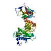
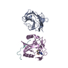
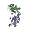
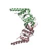
| Title | Structural analysis and inhibition of TraE from the pKM101 type IV secretion system | ||||||
 Components Components | Conjugal transfer protein | ||||||
 Keywords Keywords |  PROTEIN TRANSPORT / bacterial secretion / type IV secretion / VirB PROTEIN TRANSPORT / bacterial secretion / type IV secretion / VirB | ||||||
| Function / homology |  Function and homology information Function and homology information | ||||||
| Biological species |  Escherichia coli (E. coli) Escherichia coli (E. coli) | ||||||
| Method | X-RAY DIFFRACTION / SYNCHROTRON / MOLECULAR REPLACEMENT / Resolution: 2.441 Å | ||||||
 Authors Authors | Casu, B. / Sygusch, J. / Baron, C. | ||||||
| Funding support |  Canada, 1items Canada, 1items
| ||||||
 Citation Citation | Journal: J.Biol.Chem. / Year: 2016 Title: Structural Analysis and Inhibition of TraE from the pKM101 Type IV Secretion System. Authors: Casu, B. / Smart, J. / Hancock, M.A. / Smith, M. / Sygusch, J. / Baron, C. | ||||||
| History |
|
- Structure visualization
| Structure viewer | Molecule: MolmilJmol/JSmol |
|---|
- Downloads & links
-Download
| PDBx/mmCIF format | 5i97.cif.gz | 236.6 KB | Display | PDBx/mmCIF format |
|---|---|---|---|---|
| PDB format | pdb5i97.ent.gz | 192 KB | Display | PDB format |
| PDBx/mmJSON format | 5i97.json.gz | Tree view | PDBx/mmJSON format | |
| Others |  Other downloads Other downloads |
-Validation report
| Arichive directory | https://data.pdbj.org/pub/pdb/validation_reports/i9/5i97ftp://data.pdbj.org/pub/pdb/validation_reports/i9/5i97 | HTTPS FTP |
|---|
-Related structure data
| Related structure data |  2cc3S S: Starting model for refinement |
|---|---|
| Similar structure data |


-Links
 PDBj
PDBj
- Assembly
| Deposited unit | 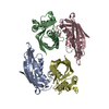
| ||||||||
|---|---|---|---|---|---|---|---|---|---|
| 1 | 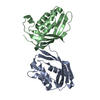
| ||||||||
| 2 | 
| ||||||||
| Unit cell |
|
-Components
| #1: Protein | Mass: 18818.104 Da / Num. of mol.: 4 / Fragment: residues 42-204 Source method: isolated from a genetically manipulated source Source: (gene. exp.) Escherichia coli (E. coli)Gene: traE, B634_00049, ECONIH1_27615, pec386IL_00125, pEcNDM0_00049, pHKU1_33, pKC394-028, pKC396-021, pMUR050_041, pN3_023, PU53_15805 Production host: Escherichia coli (E. coli) / Strain (production host): BL21(DE3)star / References: UniProt: Q17U16#2: Water | ChemComp-HOH / | Water Mass: 18.015 Da / Num. of mol.: 67 / Source method: isolated from a natural source / Formula: H2O Mass: 18.015 Da / Num. of mol.: 67 / Source method: isolated from a natural source / Formula: H2O |
|---|
-Experimental details
-Experiment
| Experiment | Method: X-RAY DIFFRACTION / Number of used crystals: 1 |
|---|
- Sample preparation
| Crystal | Density Matthews: 2.52 Å3/Da / Density % sol: 51.2 % |
|---|---|
| Crystal grow | Temperature: 293 K / Method: vapor diffusion / pH: 7.4 Details: 16% (w/v) PEG 10,000, 50 mM Bis-Tris (pH 5.5), 100 mM ammonium acetate |
-Data collection
| Diffraction | Mean temperature: 100 K |
|---|---|
| Diffraction source | Source: SYNCHROTRON / Site: NSLS  / Beamline: X25 / Wavelength: 1 Å / Beamline: X25 / Wavelength: 1 Å |
| Detector | Type: ADSC QUANTUM 315 / Detector: CCD / Date: Nov 13, 2013 |
| Radiation | Protocol: SINGLE WAVELENGTH / Monochromatic (M) / Laue (L): M / Scattering type: x-ray |
| Radiation wavelength | Wavelength: 1 Å / Relative weight: 1 |
| Reflection | Resolution: 2.441→38.6 Å / Num. obs: 28596 / % possible obs: 99.98 % / Redundancy: 6.5 % / Rmerge(I) obs: 0.121 / Net I/σ(I): 32.7 |
| Reflection shell | Resolution: 2.441→2.529 Å / Redundancy: 6 % / Mean I/σ(I) obs: 2.3 / % possible all: 99.93 |
- Processing
| Software |
| |||||||||||||||||||||||||||||||||||||||||||||||||||||||||||||||||||||||||||||||||||||||||||||||||||||||||
|---|---|---|---|---|---|---|---|---|---|---|---|---|---|---|---|---|---|---|---|---|---|---|---|---|---|---|---|---|---|---|---|---|---|---|---|---|---|---|---|---|---|---|---|---|---|---|---|---|---|---|---|---|---|---|---|---|---|---|---|---|---|---|---|---|---|---|---|---|---|---|---|---|---|---|---|---|---|---|---|---|---|---|---|---|---|---|---|---|---|---|---|---|---|---|---|---|---|---|---|---|---|---|---|---|---|---|
| Refinement | Method to determine structure: MOLECULAR REPLACEMENT Starting model: 2CC3 Resolution: 2.441→38.6 Å / SU ML: 0.32 / Cross valid method: FREE R-VALUE / σ(F): 0 / Phase error: 30.86
| |||||||||||||||||||||||||||||||||||||||||||||||||||||||||||||||||||||||||||||||||||||||||||||||||||||||||
| Solvent computation | Shrinkage radii: 0.9 Å / VDW probe radii: 1.11 Å | |||||||||||||||||||||||||||||||||||||||||||||||||||||||||||||||||||||||||||||||||||||||||||||||||||||||||
| Refinement step | Cycle: LAST / Resolution: 2.441→38.6 Å
| |||||||||||||||||||||||||||||||||||||||||||||||||||||||||||||||||||||||||||||||||||||||||||||||||||||||||
| Refine LS restraints |
| |||||||||||||||||||||||||||||||||||||||||||||||||||||||||||||||||||||||||||||||||||||||||||||||||||||||||
| LS refinement shell |
| |||||||||||||||||||||||||||||||||||||||||||||||||||||||||||||||||||||||||||||||||||||||||||||||||||||||||
| Refinement TLS params. | Method: refined / Origin x: -34.266 Å / Origin y: -19.8622 Å / Origin z: -10.2076 Å
| |||||||||||||||||||||||||||||||||||||||||||||||||||||||||||||||||||||||||||||||||||||||||||||||||||||||||
| Refinement TLS group | Selection details: all |
