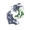
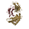
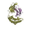
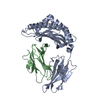
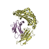
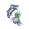
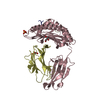
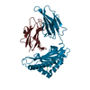
- PDB-5wes: Crystal Structure H2-Dd with disulfide-linked 5mer peptide -
+
Open data
ID or keywords:
Loading...
-
Basic information
Entry
Database: PDB / ID: 5wes
Title
Crystal Structure H2-Dd with disulfide-linked 5mer peptide
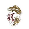
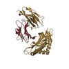
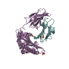
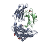
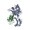
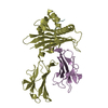
Components
Beta-2-microglobulinBeta-2 microglobulin
H-2 class I histocompatibility antigen, D-D alpha chain
Surface protein gp120
Keywords
IMMUNE SYSTEM / Antigen presentation / peptide editing / MAJOR HISTOMPATIBILITY COMPLEX CLASS I / MHC-I / TAPBPR / H2-Dd / H-2DD / TAPASIN / Peptide Loading Complex / PLC / IMMUNE RESPONSE
Function / homology
Function and homology information
Endosomal/Vacuolar pathway / DAP12 interactions / Antigen Presentation: Folding, assembly and peptide loading of class I MHC / ER-Phagosome pathway / DAP12 signaling / Immunoregulatory interactions between a Lymphoid and a non-Lymphoid cell / regulation of membrane depolarization / Dectin-2 family / antigen processing and presentation of endogenous peptide antigen via MHC class I via ER pathway, TAP-dependent / cellular defense response ...Endosomal/Vacuolar pathway / DAP12 interactions / Antigen Presentation: Folding, assembly and peptide loading of class I MHC / ER-Phagosome pathway / DAP12 signaling / Immunoregulatory interactions between a Lymphoid and a non-Lymphoid cell / regulation of membrane depolarization / Dectin-2 family / antigen processing and presentation of endogenous peptide antigen via MHC class I via ER pathway, TAP-dependent / cellular defense response / beta-2-microglobulin binding / positive regulation of plasma membrane raft polarization / positive regulation of receptor clustering / positive regulation of establishment of T cell polarity / Neutrophil degranulation / antigen processing and presentation of endogenous peptide antigen via MHC class I via ER pathway, TAP-independent / antigen processing and presentation of endogenous peptide antigen via MHC class Ib / host cell endosome membrane / lumenal side of endoplasmic reticulum membrane / peptide binding / antigen processing and presentation of exogenous protein antigen via MHC class Ib, TAP-dependent / cellular response to iron(III) ion / negative regulation of forebrain neuron differentiation / peptide antigen assembly with MHC class I protein complex / response to molecule of bacterial origin / regulation of erythrocyte differentiation / regulation of iron ion transport / MHC class I peptide loading complex / HFE-transferrin receptor complex / T cell mediated cytotoxicity / cellular response to iron ion / antigen processing and presentation of endogenous peptide antigen via MHC class I / positive regulation of T cell cytokine production / MHC class I protein complex / multicellular organismal-level iron ion homeostasis / negative regulation of neurogenesis / positive regulation of T cell mediated cytotoxicity / peptide antigen assembly with MHC class II protein complex / positive regulation of receptor-mediated endocytosis / MHC class II protein complex / cellular response to nicotine / phagocytic vesicle membrane / peptide antigen binding / positive regulation of cellular senescence / antigen processing and presentation of exogenous peptide antigen via MHC class II / negative regulation of epithelial cell proliferation / positive regulation of immune response / positive regulation of T cell activation / antimicrobial humoral immune response mediated by antimicrobial peptide / sensory perception of smell / negative regulation of neuron projection development / MHC class II protein complex binding / T cell differentiation in thymus / late endosome membrane / iron ion transport / antibacterial humoral response / protein refolding / protein homotetramerization / clathrin-dependent endocytosis of virus by host cell / intracellular iron ion homeostasis / cellular response to lipopolysaccharide / defense response to Gram-negative bacterium / amyloid fibril formation / learning or memory / viral protein processing / virus-mediated perturbation of host defense response / defense response to Gram-positive bacterium / immune response / lysosomal membrane / fusion of virus membrane with host plasma membrane / external side of plasma membrane / signaling receptor binding / innate immune response / fusion of virus membrane with host endosome membrane / viral envelope / protein-containing complex binding / virion attachment to host cell / host cell plasma membrane / virion membrane / structural molecule activity / Golgi apparatus / protein homodimerization activity / extracellular space / membrane / identical protein binding / plasma membrane / cytosol Similarity search - Function
Envelope glycoprotein Gp160 / Retroviral envelope protein / Retroviral envelope protein GP41-like / Gp120 core superfamily / Envelope glycoprotein GP120 / Human immunodeficiency virus 1, envelope glycoprotein Gp120 / MHC class I, alpha chain, C-terminal / MHC_I C-terminus / MHC class I-like antigen recognition-like / Murine Class I Major Histocompatibility Complex, H2-DB; Chain A, domain 1 ...Envelope glycoprotein Gp160 / Retroviral envelope protein / Retroviral envelope protein GP41-like / Gp120 core superfamily / Envelope glycoprotein GP120 / Human immunodeficiency virus 1, envelope glycoprotein Gp120 / MHC class I, alpha chain, C-terminal / MHC_I C-terminus / MHC class I-like antigen recognition-like / Murine Class I Major Histocompatibility Complex, H2-DB; Chain A, domain 1 / MHC class I alpha chain, alpha1 alpha2 domains / Class I Histocompatibility antigen, domains alpha 1 and 2 / Beta-2-Microglobulin / MHC class I-like antigen recognition-like / MHC class I-like antigen recognition-like superfamily / MHC classes I/II-like antigen recognition protein / Immunoglobulin/major histocompatibility complex, conserved site / Immunoglobulins and major histocompatibility complex proteins signature. / Immunoglobulin C-Type / Immunoglobulin C1-set / Immunoglobulin C1-set domain / Ig-like domain profile. / Immunoglobulin-like domain / Immunoglobulin-like domain superfamily / Immunoglobulins / Immunoglobulin-like fold / Immunoglobulin-like / Sandwich / 2-Layer Sandwich / Mainly Beta / Alpha Beta Similarity search - Domain/homology
In the structure databanks used in Yorodumi, some data are registered as the other names, "COVID-19 virus" and "2019-nCoV". Here are the details of the virus and the list of structure data.
Jan 31, 2019. EMDB accession codes are about to change! (news from PDBe EMDB page)
EMDB accession codes are about to change! (news from PDBe EMDB page)
The allocation of 4 digits for EMDB accession codes will soon come to an end. Whilst these codes will remain in use, new EMDB accession codes will include an additional digit and will expand incrementally as the available range of codes is exhausted. The current 4-digit format prefixed with “EMD-” (i.e. EMD-XXXX) will advance to a 5-digit format (i.e. EMD-XXXXX), and so on. It is currently estimated that the 4-digit codes will be depleted around Spring 2019, at which point the 5-digit format will come into force.
The EM Navigator/Yorodumi systems omit the EMD- prefix.
Related info.:Q: What is EMD? / ID/Accession-code notation in Yorodumi/EM Navigator
Yorodumi is a browser for structure data from EMDB, PDB, SASBDB, etc.
This page is also the successor to EM Navigator detail page, and also detail information page/front-end page for Omokage search.
The word "yorodu" (or yorozu) is an old Japanese word meaning "ten thousand". "mi" (miru) is to see.
Related info.:EMDB / PDB / SASBDB / Comparison of 3 databanks / Yorodumi Search / Aug 31, 2016. New EM Navigator & Yorodumi / Yorodumi Papers / Jmol/JSmol / Function and homology information / Changes in new EM Navigator and Yorodumi
 Movie
Movie Controller


 Open data
 Basic information
 Components
 Keywords
 IMMUNE SYSTEM /
  Function and homology information

 Authors
 Citation
 Structure visualization
 Downloads & links
 Other downloads


 PDBj
PDBj


 Assembly
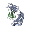


 Type: peptide linking / Mass: 75.067 Da / Num. of mol.: 1 / Source method: obtained synthetically / Formula: C2H5NO2
 Type: L-peptide linking / Mass: 131.173 Da / Num. of mol.: 1 / Source method: obtained synthetically / Formula: C6H13NO2
 Sample preparation
 / Beamline: 22-ID / Wavelength: 1.033 Å
 Processing