 Movie
Movie Controller

[English] 日本語
 Yorodumi
- PDB-5v8z: Crystal structure of ERp29 D-domain in complex with the P-domain ... -
+ Open data

- Basic information
| Entry | Database: PDB / ID: 5v8z | ||||||
|---|---|---|---|---|---|---|---|
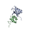
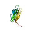
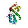
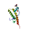
| Title | Crystal structure of ERp29 D-domain in complex with the P-domain of calmegin | ||||||
 Components Components |
| ||||||
 Keywords Keywords |  CHAPERONE / protein binding / protein folding CHAPERONE / protein binding / protein folding | ||||||
| Function / homology |  Function and homology information Function and homology informationregulation of endoplasmic reticulum stress-induced intrinsic apoptotic signaling pathway / smooth endoplasmic reticulum / : / protein secretion / protein unfolding / negative regulation of protein secretion / transport vesicle / intracellular protein transport / positive regulation of MAP kinase activity / unfolded protein binding ...regulation of endoplasmic reticulum stress-induced intrinsic apoptotic signaling pathway / smooth endoplasmic reticulum / : / protein secretion / protein unfolding / negative regulation of protein secretion / transport vesicle / intracellular protein transport / positive regulation of MAP kinase activity / unfolded protein binding / melanosome / protein folding / protein-folding chaperone binding / positive regulation of protein phosphorylation / endoplasmic reticulum lumen / negative regulation of gene expression / calcium ion binding / endoplasmic reticulum membrane / positive regulation of gene expression / cell surface / endoplasmic reticulum / protein homodimerization activity / membraneSimilarity search - Function | ||||||
| Biological species |  Homo sapiens (human) Homo sapiens (human) Canis lupus familiaris (dog) Canis lupus familiaris (dog) | ||||||
| Method | X-RAY DIFFRACTION / SYNCHROTRON / MOLECULAR REPLACEMENT / Resolution: 2.105 Å | ||||||
 Authors Authors | Kozlov, G. / Munoz-Escobar, J. / Gehring, K. | ||||||
| Funding support |  Canada, 1items Canada, 1items
| ||||||
 Citation Citation | Journal: Structure / Year: 2017 Title: Mapping the ER Interactome: The P Domains of Calnexin and Calreticulin as Plurivalent Adapters for Foldases and Chaperones. Authors: Kozlov, G. / Munoz-Escobar, J. / Castro, K. / Gehring, K. | ||||||
| History |
|
- Structure visualization
| Structure viewer | Molecule: MolmilJmol/JSmol |
|---|
- Downloads & links
-Download
| PDBx/mmCIF format | 5v8z.cif.gz | 63.6 KB | Display | PDBx/mmCIF format |
|---|---|---|---|---|
| PDB format | pdb5v8z.ent.gz | 46 KB | Display | PDB format |
| PDBx/mmJSON format | 5v8z.json.gz | Tree view | PDBx/mmJSON format | |
| Others |  Other downloads Other downloads |
-Validation report
| Arichive directory | https://data.pdbj.org/pub/pdb/validation_reports/v8/5v8zftp://data.pdbj.org/pub/pdb/validation_reports/v8/5v8z | HTTPS FTP |
|---|
-Related structure data
| Related structure data |  5v90C  2qc7S C: citing same article ( S: Starting model for refinement |
|---|---|
| Similar structure data |


-Links
 PDBj
PDBj


- Assembly
| Deposited unit | 
| ||||||||
|---|---|---|---|---|---|---|---|---|---|
| 1 | 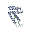
| ||||||||
| 2 | 
| ||||||||
| Unit cell |
|
-Components
| #1: Protein | Mass: 11903.626 Da / Num. of mol.: 2 / Fragment: UNP residues 158-261 Source method: isolated from a genetically manipulated source Source: (gene. exp.) Homo sapiens (human) / Gene: ERP29, C12orf8, ERP28 / Production host:  Escherichia coli (E. coli) / References: UniProt: P30040 Escherichia coli (E. coli) / References: UniProt: P30040#2: Protein/peptide | Mass: 4342.601 Da / Num. of mol.: 2 / Fragment: UNP residues 327-360 Source method: isolated from a genetically manipulated source Source: (gene. exp.) Canis lupus familiaris (dog) / Gene: CLGN / Plasmid: pET15b / Production host: Escherichia coli (E. coli) / References: UniProt: E2RA18#3: Water | ChemComp-HOH / | Water Mass: 18.015 Da / Num. of mol.: 37 / Source method: isolated from a natural source / Formula: H2O Mass: 18.015 Da / Num. of mol.: 37 / Source method: isolated from a natural source / Formula: H2O |
|---|
-Experimental details
-Experiment
| Experiment | Method: X-RAY DIFFRACTION / Number of used crystals: 1 |
|---|
- Sample preparation
| Crystal | Density Matthews: 2.59 Å3/Da / Density % sol: 52.54 % |
|---|---|
| Crystal grow | Temperature: 293 K / Method: vapor diffusion, sitting drop / pH: 7.5 / Details: 1.4 M sodium citrate pH 7.5 |
-Data collection
| Diffraction | Mean temperature: 100 K |
|---|---|
| Diffraction source | Source: SYNCHROTRON / Site: CHESS  / Beamline: F1 / Wavelength: 0.9782 Å / Beamline: F1 / Wavelength: 0.9782 Å |
| Detector | Type: DECTRIS PILATUS3 6M / Detector: PIXEL / Date: Nov 11, 2015 |
| Radiation | Protocol: SINGLE WAVELENGTH / Monochromatic (M) / Laue (L): M / Scattering type: x-ray |
| Radiation wavelength | Wavelength: 0.9782 Å / Relative weight: 1 |
| Reflection | Resolution: 2.1→50 Å / Num. obs: 20065 / % possible obs: 99.7 % / Redundancy: 8.6 % / Rsym value: 0.087 / Net I/σ(I): 34.4 |
| Reflection shell | Resolution: 2.1→2.14 Å / Redundancy: 9 % / Mean I/σ(I) obs: 3.2 / Num. unique all: 1948 / Num. unique obs: 1021 / Rsym value: 0.512 / % possible all: 99 |
- Processing
| Software |
| ||||||||||||||||||||||||||||||||||||||||||||||||||||||||
|---|---|---|---|---|---|---|---|---|---|---|---|---|---|---|---|---|---|---|---|---|---|---|---|---|---|---|---|---|---|---|---|---|---|---|---|---|---|---|---|---|---|---|---|---|---|---|---|---|---|---|---|---|---|---|---|---|---|
| Refinement | Method to determine structure: MOLECULAR REPLACEMENT Starting model: 2QC7 Resolution: 2.105→33.81 Å / SU ML: 0.21 / Cross valid method: THROUGHOUT / σ(F): 1.38 / Phase error: 25.49
| ||||||||||||||||||||||||||||||||||||||||||||||||||||||||
| Solvent computation | Shrinkage radii: 0.9 Å / VDW probe radii: 1.11 Å | ||||||||||||||||||||||||||||||||||||||||||||||||||||||||
| Refinement step | Cycle: LAST / Resolution: 2.105→33.81 Å
| ||||||||||||||||||||||||||||||||||||||||||||||||||||||||
| Refine LS restraints |
| ||||||||||||||||||||||||||||||||||||||||||||||||||||||||
| LS refinement shell |
|
