 Movie
Movie Controller


+ Open data

- Basic information
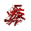
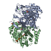
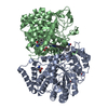
| Entry | Database: PDB / ID: 5uxn | ||||||
|---|---|---|---|---|---|---|---|
| Title | Type II DAH7PS from Pseudomonas aeruginosa with Tyr bound | ||||||
 Components Components | Phospho-2-dehydro-3-deoxyheptonate aldolase | ||||||
 Keywords Keywords |  TRANSFERASE / DAH7PS / 3-deoxy-D-arabino-heptulosonate 7-phosphate synthase / shikimate pathway TRANSFERASE / DAH7PS / 3-deoxy-D-arabino-heptulosonate 7-phosphate synthase / shikimate pathway | ||||||
| Function / homology |  Function and homology information3-deoxy-7-phosphoheptulonate synthase / 3-deoxy-7-phosphoheptulonate synthase activity / aromatic amino acid family biosynthetic process Function and homology information3-deoxy-7-phosphoheptulonate synthase / 3-deoxy-7-phosphoheptulonate synthase activity / aromatic amino acid family biosynthetic processSimilarity search - Function | ||||||
| Biological species |   Pseudomonas aeruginosa (bacteria) Pseudomonas aeruginosa (bacteria) | ||||||
| Method | X-RAY DIFFRACTION / SYNCHROTRON / Resolution: 2.2 Å | ||||||
 Authors Authors | Sterritt, O.W. / Jameson, G.B. / Parker, E.J. | ||||||
 Citation Citation | Journal: Biochemistry / Year: 2018 Title: A Pseudoisostructural Type II DAH7PS Enzyme from Pseudomonas aeruginosa: Alternative Evolutionary Strategies to Control Shikimate Pathway Flux. Authors: Sterritt, O.W. / Kessans, S.A. / Jameson, G.B. / Parker, E.J. | ||||||
| History |
|
- Structure visualization
| Structure viewer | Molecule: MolmilJmol/JSmol |
|---|
- Downloads & links
-Download
| PDBx/mmCIF format | 5uxn.cif.gz | 105.3 KB | Display | PDBx/mmCIF format |
|---|---|---|---|---|
| PDB format | pdb5uxn.ent.gz | 77.9 KB | Display | PDB format |
| PDBx/mmJSON format | 5uxn.json.gz | Tree view | PDBx/mmJSON format | |
| Others |  Other downloads Other downloads |
-Validation report
| Arichive directory | https://data.pdbj.org/pub/pdb/validation_reports/ux/5uxnftp://data.pdbj.org/pub/pdb/validation_reports/ux/5uxn | HTTPS FTP |
|---|
-Related structure data


-Links
 PDBj
PDBj


- Assembly
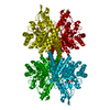
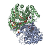
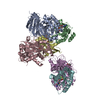
| Deposited unit | 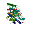
| ||||||||
|---|---|---|---|---|---|---|---|---|---|
| 1 | 
| ||||||||
| Unit cell |
| ||||||||
| Components on special symmetry positions |
|
-Components
-Protein , 1 types, 1 molecules A
| #1: Protein | Mass: 50197.375 Da / Num. of mol.: 1 Source method: isolated from a genetically manipulated source Source: (gene. exp.) Pseudomonas aeruginosa (strain ATCC 15692 / DSM 22644 / CIP 104116 / JCM 14847 / LMG 12228 / 1C / PRS 101 / PAO1) (bacteria)Strain: ATCC 15692 / DSM 22644 / CIP 104116 / JCM 14847 / LMG 12228 / 1C / PRS 101 / PAO1 Gene: PA2843 / Plasmid: pET28a / Details (production host): N-terminal His tag + linker / Production host: Escherichia coli (E. coli) / Strain (production host): BL21(DE3)References: UniProt: Q9I000, 3-deoxy-7-phosphoheptulonate synthase |
|---|
-Non-polymers , 6 types, 148 molecules 

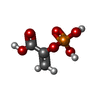


| #2: Chemical | Phosphate Mass: 94.971 Da / Num. of mol.: 2 / Source method: obtained synthetically / Formula: PO4 Mass: 94.971 Da / Num. of mol.: 2 / Source method: obtained synthetically / Formula: PO4#3: Chemical | ChemComp-CO / |  Mass: 58.933 Da / Num. of mol.: 1 / Source method: obtained synthetically / Formula: Co Mass: 58.933 Da / Num. of mol.: 1 / Source method: obtained synthetically / Formula: Co#4: Chemical | ChemComp-PEP / | Phosphoenolpyruvic acid Mass: 168.042 Da / Num. of mol.: 1 / Source method: obtained synthetically / Formula: C3H5O6P Mass: 168.042 Da / Num. of mol.: 1 / Source method: obtained synthetically / Formula: C3H5O6P#5: Chemical | ChemComp-TYR / | Tyrosine Type: L-peptide linking / Mass: 181.189 Da / Num. of mol.: 1 / Source method: obtained synthetically / Formula: C9H11NO3 Type: L-peptide linking / Mass: 181.189 Da / Num. of mol.: 1 / Source method: obtained synthetically / Formula: C9H11NO3#6: Chemical | ChemComp-CL / | Chloride Mass: 35.453 Da / Num. of mol.: 1 / Source method: obtained synthetically / Formula: Cl Mass: 35.453 Da / Num. of mol.: 1 / Source method: obtained synthetically / Formula: Cl#7: Water | ChemComp-HOH / | WaterMass: 18.015 Da / Num. of mol.: 142 / Source method: isolated from a natural source / Formula: H2O |
|---|
-Experimental details
-Experiment
| Experiment | Method: X-RAY DIFFRACTION / Number of used crystals: 1 |
|---|
- Sample preparation
| Crystal | Density Matthews: 2.35 Å3/Da / Density % sol: 47.68 % / Description: rod |
|---|---|
| Crystal grow | Temperature: 278 K / Method: vapor diffusion, hanging drop / pH: 5.5 Details: 0.1 M sodium acetate pH 5.5, 0.8 M sodium formate, 1 mM cobalt chloride, 1 mM phosphoenol pyruvate, 3 mM Tyr, 26 % PEG 2000 MME Temp details: Temperature controlled |
-Data collection
| Diffraction | Mean temperature: 80 K |
|---|---|
| Diffraction source | Source: SYNCHROTRON / Site: Australian Synchrotron  / Beamline: MX1 / Wavelength: 0.9537 Å / Beamline: MX1 / Wavelength: 0.9537 Å |
| Detector | Type: ADSC QUANTUM 315r / Detector: CCD / Date: Aug 5, 2016 |
| Radiation | Monochromator: Double Crystal SI(111) water cooled / Protocol: SINGLE WAVELENGTH / Monochromatic (M) / Laue (L): M / Scattering type: x-ray |
| Radiation wavelength | Wavelength: 0.9537 Å / Relative weight: 1 |
| Reflection | Resolution: 2.2→57.63 Å / Num. obs: 24329 / % possible obs: 100 % / Redundancy: 7.1 % / Biso Wilson estimate: 13.3 Å2 / CC1/2: 0.992 / Rmerge(I) obs: 0.139 / Rpim(I) all: 0.056 / Net I/σ(I): 10.5 |
| Reflection shell | Resolution: 2.2→2.27 Å / Redundancy: 7.3 % / Rmerge(I) obs: 0.326 / Mean I/σ(I) obs: 6.4 / Num. unique obs: 2093 / CC1/2: 0.936 / Rpim(I) all: 0.13 / % possible all: 100 |
- Processing
| Software |
| ||||||||||||||||||||||||||||||||||||||||||||||||||||||||||||||||||||||||||||||||||||||||||||||||||||||||||||||||||||||||||||||||||||||||||||||||||||||||||||||||||||||||||||||||||||||
|---|---|---|---|---|---|---|---|---|---|---|---|---|---|---|---|---|---|---|---|---|---|---|---|---|---|---|---|---|---|---|---|---|---|---|---|---|---|---|---|---|---|---|---|---|---|---|---|---|---|---|---|---|---|---|---|---|---|---|---|---|---|---|---|---|---|---|---|---|---|---|---|---|---|---|---|---|---|---|---|---|---|---|---|---|---|---|---|---|---|---|---|---|---|---|---|---|---|---|---|---|---|---|---|---|---|---|---|---|---|---|---|---|---|---|---|---|---|---|---|---|---|---|---|---|---|---|---|---|---|---|---|---|---|---|---|---|---|---|---|---|---|---|---|---|---|---|---|---|---|---|---|---|---|---|---|---|---|---|---|---|---|---|---|---|---|---|---|---|---|---|---|---|---|---|---|---|---|---|---|---|---|---|---|
| Refinement | Resolution: 2.2→57.63 Å / Cor.coef. Fo:Fc: 0.941 / Cor.coef. Fo:Fc free: 0.902 / SU B: 5.313 / SU ML: 0.139 / Cross valid method: THROUGHOUT / ESU R: 0.263 / ESU R Free: 0.2 / Details: HYDROGENS HAVE BEEN ADDED IN THE RIDING POSITIONS
| ||||||||||||||||||||||||||||||||||||||||||||||||||||||||||||||||||||||||||||||||||||||||||||||||||||||||||||||||||||||||||||||||||||||||||||||||||||||||||||||||||||||||||||||||||||||
| Solvent computation | Ion probe radii: 0.8 Å / Shrinkage radii: 0.8 Å / VDW probe radii: 1.2 Å | ||||||||||||||||||||||||||||||||||||||||||||||||||||||||||||||||||||||||||||||||||||||||||||||||||||||||||||||||||||||||||||||||||||||||||||||||||||||||||||||||||||||||||||||||||||||
| Displacement parameters | Biso mean: 23.87 Å2
| ||||||||||||||||||||||||||||||||||||||||||||||||||||||||||||||||||||||||||||||||||||||||||||||||||||||||||||||||||||||||||||||||||||||||||||||||||||||||||||||||||||||||||||||||||||||
| Refinement step | Cycle: 1 / Resolution: 2.2→57.63 Å
| ||||||||||||||||||||||||||||||||||||||||||||||||||||||||||||||||||||||||||||||||||||||||||||||||||||||||||||||||||||||||||||||||||||||||||||||||||||||||||||||||||||||||||||||||||||||
| Refine LS restraints |
|