 Movie
Movie Controller

[English] 日本語
 Yorodumi
- PDB-5uv4: Crystal Structure of Maize SIRK1 (sucrose-induced receptor kinase... -
+ Open data

- Basic information
| Entry | Database: PDB / ID: 5uv4 | ||||||
|---|---|---|---|---|---|---|---|
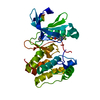
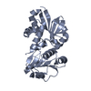
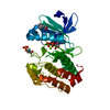
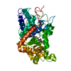
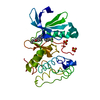
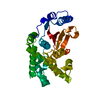
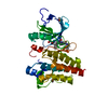
| Title | Crystal Structure of Maize SIRK1 (sucrose-induced receptor kinase 1) kinase domain bound to AMP-PNP | ||||||
 Components Components | Putative leucine-rich repeat protein kinase family protein | ||||||
 Keywords Keywords | TRANSFERASE / receptor-like kinase / leucine-rich repeat / sucrose-induced kinase / Structural Genomics / Structural Genomics Consortium / SGC | ||||||
| Function / homology |  Function and homology information Function and homology information | ||||||
| Biological species |  Zea mays (maize) Zea mays (maize) | ||||||
| Method | X-RAY DIFFRACTION / SYNCHROTRON / MOLECULAR REPLACEMENT / Resolution: 2.3 Å | ||||||
 Authors Authors | Counago, R.M. / Aquino, B. / Massirer, K.B. / Gileadi, O. / Arruda, P. / Structural Genomics Consortium (SGC) | ||||||
| Funding support |  Brazil, 1items Brazil, 1items
| ||||||
 Citation Citation | Journal: To Be Published Title: Crystal Structure of Maize SIRK1 (sucrose-induced receptor kinase 1) kinase domain bound to AMP-PNP Authors: Aquino, B. / Counago, R.M. / Massirer, K.B. / Gileadi, O. / Arruda, P. / Structural Genomics Consortium (SGC) | ||||||
| History |
|
- Structure visualization
| Structure viewer | Molecule: MolmilJmol/JSmol |
|---|
- Downloads & links
-Download
| PDBx/mmCIF format | 5uv4.cif.gz | 131.5 KB | Display | PDBx/mmCIF format |
|---|---|---|---|---|
| PDB format | pdb5uv4.ent.gz | 99.4 KB | Display | PDB format |
| PDBx/mmJSON format | 5uv4.json.gz | Tree view | PDBx/mmJSON format | |
| Others |  Other downloads Other downloads |
-Validation report
| Arichive directory | https://data.pdbj.org/pub/pdb/validation_reports/uv/5uv4ftp://data.pdbj.org/pub/pdb/validation_reports/uv/5uv4 | HTTPS FTP |
|---|
-Related structure data
| Related structure data |  4l68S S: Starting model for refinement |
|---|---|
| Similar structure data |


-Links
 PDBj
PDBj


- Assembly
| Deposited unit | 
| ||||||||
|---|---|---|---|---|---|---|---|---|---|
| 1 |
| ||||||||
| Unit cell |
|
-Components
| #1: Protein | Mass: 34498.344 Da / Num. of mol.: 1 / Fragment: UNP residues 737-1045 Source method: isolated from a genetically manipulated source Source: (gene. exp.) Zea mays (maize) / Gene: ZEAMMB73_708518 / Production host:  Escherichia coli (E. coli) / Strain (production host): BL21 DE3 R3 / References: UniProt: K7VIQ3 Escherichia coli (E. coli) / Strain (production host): BL21 DE3 R3 / References: UniProt: K7VIQ3 |
|---|---|
| #2: Chemical | ChemComp-ANP /   Mass: 506.196 Da / Num. of mol.: 1 / Source method: obtained synthetically / Formula: C10H17N6O12P3 / Comment: AMP-PNP, energy-carrying molecule analogue*YM Mass: 506.196 Da / Num. of mol.: 1 / Source method: obtained synthetically / Formula: C10H17N6O12P3 / Comment: AMP-PNP, energy-carrying molecule analogue*YM |
| #3: Chemical | ChemComp-MG /   Mass: 24.305 Da / Num. of mol.: 1 / Source method: obtained synthetically / Formula: Mg Mass: 24.305 Da / Num. of mol.: 1 / Source method: obtained synthetically / Formula: Mg |
| #4: Water | ChemComp-HOH / Water Mass: 18.015 Da / Num. of mol.: 65 / Source method: isolated from a natural source / Formula: H2O Mass: 18.015 Da / Num. of mol.: 65 / Source method: isolated from a natural source / Formula: H2O |
-Experimental details
-Experiment
| Experiment | Method: X-RAY DIFFRACTION / Number of used crystals: 1 |
|---|
- Sample preparation
| Crystal | Density Matthews: 2.3 Å3/Da / Density % sol: 46.56 % |
|---|---|
| Crystal grow | Temperature: 293 K / Method: vapor diffusion, sitting drop Details: 12.5% w/v PEG1000, 12.5% w/v PEG3350, 12.5% v/v MPD, 0,02M of each monossacharide (D-glucose, D-mannose, D-galactose, L-fucose, D-xylose, N-acetyl-D-glucosamine), 0.1M MOPS/HEPES-Na pH 7.5 |
-Data collection
| Diffraction | Mean temperature: 100 K |
|---|---|
| Diffraction source | Source: SYNCHROTRON / Site: APS  / Beamline: 24-ID-C / Wavelength: 0.9792 Å / Beamline: 24-ID-C / Wavelength: 0.9792 Å |
| Detector | Type: DECTRIS PILATUS 6M-F / Detector: PIXEL / Date: Nov 9, 2016 |
| Radiation | Monochromator: Si(111) / Protocol: SINGLE WAVELENGTH / Monochromatic (M) / Laue (L): M / Scattering type: x-ray |
| Radiation wavelength | Wavelength: 0.9792 Å / Relative weight: 1 |
| Reflection | Resolution: 2.3→39.87 Å / Num. obs: 14190 / % possible obs: 97 % / Redundancy: 4 % / Biso Wilson estimate: 55.34 Å2 / CC1/2: 0.998 / Rmerge(I) obs: 0.049 / Rpim(I) all: 0.027 / Net I/σ(I): 14.8 |
| Reflection shell | Resolution: 2.3→2.39 Å / Redundancy: 4.1 % / Rmerge(I) obs: 0.551 / Mean I/σ(I) obs: 2.6 / Num. unique all: 1492 / Num. unique obs: 1492 / CC1/2: 0.905 / Rpim(I) all: 0.341 / % possible all: 98.4 |
- Processing
| Software |
| |||||||||||||||||||||||||||||||||||||||||||||||||||||||||||||||||||||||||||||||||||||||||||||||||||||||||||||||||||||||||||||||||||||||||||||||||||||||||||||||||||||||||||||||
|---|---|---|---|---|---|---|---|---|---|---|---|---|---|---|---|---|---|---|---|---|---|---|---|---|---|---|---|---|---|---|---|---|---|---|---|---|---|---|---|---|---|---|---|---|---|---|---|---|---|---|---|---|---|---|---|---|---|---|---|---|---|---|---|---|---|---|---|---|---|---|---|---|---|---|---|---|---|---|---|---|---|---|---|---|---|---|---|---|---|---|---|---|---|---|---|---|---|---|---|---|---|---|---|---|---|---|---|---|---|---|---|---|---|---|---|---|---|---|---|---|---|---|---|---|---|---|---|---|---|---|---|---|---|---|---|---|---|---|---|---|---|---|---|---|---|---|---|---|---|---|---|---|---|---|---|---|---|---|---|---|---|---|---|---|---|---|---|---|---|---|---|---|---|---|---|---|
| Refinement | Method to determine structure: MOLECULAR REPLACEMENT Starting model: 4L68 Resolution: 2.3→39.87 Å / Cor.coef. Fo:Fc: 0.929 / Cor.coef. Fo:Fc free: 0.913 / SU R Cruickshank DPI: 0.284 / Cross valid method: THROUGHOUT / σ(F): 0 / SU R Blow DPI: 0.296 / SU Rfree Blow DPI: 0.197 / SU Rfree Cruickshank DPI: 0.196
| |||||||||||||||||||||||||||||||||||||||||||||||||||||||||||||||||||||||||||||||||||||||||||||||||||||||||||||||||||||||||||||||||||||||||||||||||||||||||||||||||||||||||||||||
| Displacement parameters | Biso mean: 66.92 Å2
| |||||||||||||||||||||||||||||||||||||||||||||||||||||||||||||||||||||||||||||||||||||||||||||||||||||||||||||||||||||||||||||||||||||||||||||||||||||||||||||||||||||||||||||||
| Refinement step | Cycle: 1 / Resolution: 2.3→39.87 Å
| |||||||||||||||||||||||||||||||||||||||||||||||||||||||||||||||||||||||||||||||||||||||||||||||||||||||||||||||||||||||||||||||||||||||||||||||||||||||||||||||||||||||||||||||
| Refine LS restraints |
| |||||||||||||||||||||||||||||||||||||||||||||||||||||||||||||||||||||||||||||||||||||||||||||||||||||||||||||||||||||||||||||||||||||||||||||||||||||||||||||||||||||||||||||||
| LS refinement shell | Resolution: 2.3→2.48 Å / Total num. of bins used: 7
| |||||||||||||||||||||||||||||||||||||||||||||||||||||||||||||||||||||||||||||||||||||||||||||||||||||||||||||||||||||||||||||||||||||||||||||||||||||||||||||||||||||||||||||||
| Refinement TLS params. | Method: refined / Refine-ID: X-RAY DIFFRACTION
| |||||||||||||||||||||||||||||||||||||||||||||||||||||||||||||||||||||||||||||||||||||||||||||||||||||||||||||||||||||||||||||||||||||||||||||||||||||||||||||||||||||||||||||||
| Refinement TLS group |
|
