 Movie
Movie Controller

[English] 日本語
 Yorodumi
- PDB-5upe: Crystal structure of human NAMPT with isoindoline urea inhibitor ... -
+ Open data

- Basic information
| Entry | Database: PDB / ID: 5upe | ||||||
|---|---|---|---|---|---|---|---|
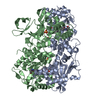
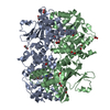
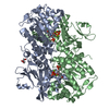
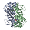
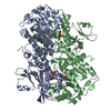
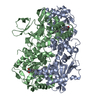
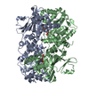
| Title | Crystal structure of human NAMPT with isoindoline urea inhibitor compound 5 | ||||||
 Components Components | Nicotinamide phosphoribosyltransferase | ||||||
 Keywords Keywords | TRANSFERASE/TRANSFERASE INHIBITOR / NAMPT inhibitor / TRANSFERASE-TRANSFERASE INHIBITOR complex | ||||||
| Function / homology |  Function and homology informationnicotinamide phosphoribosyltransferase / nicotinamide phosphoribosyltransferase activity / NAD biosynthesis via nicotinamide riboside salvage pathway / Nicotinamide salvaging / NAD biosynthetic process / positive regulation of nitric-oxide synthase biosynthetic process / NPAS4 regulates expression of target genes / BMAL1:CLOCK,NPAS2 activates circadian gene expression / cytokine activity / circadian regulation of gene expression ...nicotinamide phosphoribosyltransferase / nicotinamide phosphoribosyltransferase activity / NAD biosynthesis via nicotinamide riboside salvage pathway / Nicotinamide salvaging / NAD biosynthetic process / positive regulation of nitric-oxide synthase biosynthetic process / NPAS4 regulates expression of target genes / BMAL1:CLOCK,NPAS2 activates circadian gene expression / cytokine activity / circadian regulation of gene expression / cell junction / cell-cell signaling / nuclear speck / positive regulation of cell population proliferation / signal transduction / positive regulation of transcription by RNA polymerase II / extracellular exosome / identical protein binding / cytosol Function and homology informationnicotinamide phosphoribosyltransferase / nicotinamide phosphoribosyltransferase activity / NAD biosynthesis via nicotinamide riboside salvage pathway / Nicotinamide salvaging / NAD biosynthetic process / positive regulation of nitric-oxide synthase biosynthetic process / NPAS4 regulates expression of target genes / BMAL1:CLOCK,NPAS2 activates circadian gene expression / cytokine activity / circadian regulation of gene expression ...nicotinamide phosphoribosyltransferase / nicotinamide phosphoribosyltransferase activity / NAD biosynthesis via nicotinamide riboside salvage pathway / Nicotinamide salvaging / NAD biosynthetic process / positive regulation of nitric-oxide synthase biosynthetic process / NPAS4 regulates expression of target genes / BMAL1:CLOCK,NPAS2 activates circadian gene expression / cytokine activity / circadian regulation of gene expression / cell junction / cell-cell signaling / nuclear speck / positive regulation of cell population proliferation / signal transduction / positive regulation of transcription by RNA polymerase II / extracellular exosome / identical protein binding / cytosolSimilarity search - Function | ||||||
| Biological species |  Homo sapiens (human) Homo sapiens (human) | ||||||
| Method | X-RAY DIFFRACTION / SYNCHROTRON / MOLECULAR REPLACEMENT / Resolution: 1.93 Å | ||||||
 Authors Authors | Longenecker, K.L. / Raich, D. / Korepanova, A.V. | ||||||
 Citation Citation | Journal: Bioorg. Med. Chem. Lett. / Year: 2017 Title: SAR and characterization of non-substrate isoindoline urea inhibitors of nicotinamide phosphoribosyltransferase (NAMPT). Authors: Curtin, M.L. / Heyman, H.R. / Clark, R.F. / Sorensen, B.K. / Doherty, G.A. / Hansen, T.M. / Frey, R.R. / Sarris, K.A. / Aguirre, A.L. / Shrestha, A. / Tu, N. / Woller, K. / Pliushchev, M.A. ...Authors: Curtin, M.L. / Heyman, H.R. / Clark, R.F. / Sorensen, B.K. / Doherty, G.A. / Hansen, T.M. / Frey, R.R. / Sarris, K.A. / Aguirre, A.L. / Shrestha, A. / Tu, N. / Woller, K. / Pliushchev, M.A. / Sweis, R.F. / Cheng, M. / Wilsbacher, J.L. / Kovar, P.J. / Guo, J. / Cheng, D. / Longenecker, K.L. / Raich, D. / Korepanova, A.V. / Soni, N.B. / Algire, M.A. / Richardson, P.L. / Marin, V.L. / Badagnani, I. / Vasudevan, A. / Buchanan, F.G. / Maag, D. / Chiang, G.G. / Tse, C. / Michaelides, M.R. | ||||||
| History |
|
- Structure visualization
| Structure viewer | Molecule: MolmilJmol/JSmol |
|---|
- Downloads & links
-Download
| PDBx/mmCIF format | 5upe.cif.gz | 210.7 KB | Display | PDBx/mmCIF format |
|---|---|---|---|---|
| PDB format | pdb5upe.ent.gz | 165.3 KB | Display | PDB format |
| PDBx/mmJSON format | 5upe.json.gz | Tree view | PDBx/mmJSON format | |
| Others |  Other downloads Other downloads |
-Validation report
| Arichive directory | https://data.pdbj.org/pub/pdb/validation_reports/up/5upeftp://data.pdbj.org/pub/pdb/validation_reports/up/5upe | HTTPS FTP |
|---|
-Related structure data
| Related structure data |  5upfC 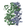 2gvjS C: citing same article ( S: Starting model for refinement |
|---|---|
| Similar structure data |


-Links
 PDBj
PDBj- Assembly
| Deposited unit | 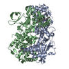
| ||||||||
|---|---|---|---|---|---|---|---|---|---|
| 1 |
| ||||||||
| Unit cell |
|
-Components
| #1: Protein | / Nampt / Pre-B-cell colony-enhancing factor 1 / Pre-B cell-enhancing factor / Visfatin Mass: 56591.898 Da / Num. of mol.: 2 Source method: isolated from a genetically manipulated source Source: (gene. exp.) Homo sapiens (human) / Gene: NAMPT, PBEF, PBEF1 / Cell line (production host): HEK 293-6E / Production host: Mammalia (mammals)References: UniProt: P43490, nicotinamide phosphoribosyltransferase#2: Chemical |   Mass: 397.469 Da / Num. of mol.: 2 / Source method: obtained synthetically / Formula: C25H23N3O2 Mass: 397.469 Da / Num. of mol.: 2 / Source method: obtained synthetically / Formula: C25H23N3O2#3: Water | ChemComp-HOH / | Water Mass: 18.015 Da / Num. of mol.: 617 / Source method: isolated from a natural source / Formula: H2O Mass: 18.015 Da / Num. of mol.: 617 / Source method: isolated from a natural source / Formula: H2O |
|---|
-Experimental details
-Experiment
| Experiment | Method: X-RAY DIFFRACTION / Number of used crystals: 1 |
|---|
- Sample preparation
| Crystal | Density Matthews: 2.37 Å3/Da / Density % sol: 48.12 % |
|---|---|
| Crystal grow | Temperature: 277 K / Method: vapor diffusion, hanging drop Details: 25% PEG 3350, 0.2 M ammonium sulfate, and 0.1 M HEPES, pH 7.5 |
-Data collection
| Diffraction | Mean temperature: 100 K |
|---|---|
| Diffraction source | Source: SYNCHROTRON / Site: APS  / Beamline: 17-ID / Wavelength: 1 Å / Beamline: 17-ID / Wavelength: 1 Å |
| Detector | Type: DECTRIS PILATUS 6M / Detector: PIXEL / Date: 2010 |
| Radiation | Protocol: SINGLE WAVELENGTH / Monochromatic (M) / Laue (L): M / Scattering type: x-ray |
| Radiation wavelength | Wavelength: 1 Å / Relative weight: 1 |
| Reflection | Resolution: 1.93→82.55 Å / Num. obs: 78276 / % possible obs: 99.3 % / Redundancy: 3.3 % / Biso Wilson estimate: 25.61 Å2 / CC1/2: 0.995 / Rmerge(I) obs: 0.102 / Rpim(I) all: 0.066 / Rrim(I) all: 0.122 / Rsym value: 0.102 / Net I/σ(I): 8.9 |
| Reflection shell | Resolution: 1.933→1.94 Å / Redundancy: 3.4 % / Rmerge(I) obs: 0.865 / Mean I/σ(I) obs: 1.5 / CC1/2: 0.84 / Rpim(I) all: 0.417 / Rrim(I) all: 0.787 / Rsym value: 0.865 / % possible all: 97.7 |
- Processing
| Software |
| ||||||||||||||||||||||||||||||||||||||||||||||||||||||||||||||||||||||||||||||||||||||||||||||||||||||||||||
|---|---|---|---|---|---|---|---|---|---|---|---|---|---|---|---|---|---|---|---|---|---|---|---|---|---|---|---|---|---|---|---|---|---|---|---|---|---|---|---|---|---|---|---|---|---|---|---|---|---|---|---|---|---|---|---|---|---|---|---|---|---|---|---|---|---|---|---|---|---|---|---|---|---|---|---|---|---|---|---|---|---|---|---|---|---|---|---|---|---|---|---|---|---|---|---|---|---|---|---|---|---|---|---|---|---|---|---|---|---|
| Refinement | Method to determine structure: MOLECULAR REPLACEMENT Starting model: 2GVJ Resolution: 1.93→28.26 Å / Cor.coef. Fo:Fc: 0.923 / Cor.coef. Fo:Fc free: 0.916 / SU R Cruickshank DPI: 0.153 / Cross valid method: THROUGHOUT / σ(F): 0 / SU R Blow DPI: 0.154 / SU Rfree Blow DPI: 0.135 / SU Rfree Cruickshank DPI: 0.135
| ||||||||||||||||||||||||||||||||||||||||||||||||||||||||||||||||||||||||||||||||||||||||||||||||||||||||||||
| Displacement parameters | Biso max: 97.32 Å2 / Biso mean: 29.33 Å2 / Biso min: 11.52 Å2
| ||||||||||||||||||||||||||||||||||||||||||||||||||||||||||||||||||||||||||||||||||||||||||||||||||||||||||||
| Refine analyze | Luzzati coordinate error obs: 0.25 Å | ||||||||||||||||||||||||||||||||||||||||||||||||||||||||||||||||||||||||||||||||||||||||||||||||||||||||||||
| Refinement step | Cycle: final / Resolution: 1.93→28.26 Å
| ||||||||||||||||||||||||||||||||||||||||||||||||||||||||||||||||||||||||||||||||||||||||||||||||||||||||||||
| Refine LS restraints |
| ||||||||||||||||||||||||||||||||||||||||||||||||||||||||||||||||||||||||||||||||||||||||||||||||||||||||||||
| LS refinement shell | Resolution: 1.93→1.98 Å / Rfactor Rfree error: 0 / Total num. of bins used: 20
|
