 Movie
Movie Controller

[English] 日本語
 Yorodumi
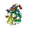
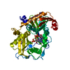
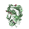
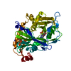
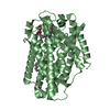
- PDB-5uli: Crystal Structure of mouse DXO in complex with (3'-NADP)+ and cal... -
+ Open data

- Basic information
| Entry | Database: PDB / ID: 5uli | ||||||
|---|---|---|---|---|---|---|---|
| Title | Crystal Structure of mouse DXO in complex with (3'-NADP)+ and calcium ion | ||||||
 Components Components | Decapping and exoribonuclease protein | ||||||
 Keywords Keywords |  HYDROLASE / NADP HYDROLASE / NADP | ||||||
| Function / homology |  Function and homology information Function and homology informationRNA NAD+-cap (NAD+-forming) hydrolase activity / mRNA 5'-diphosphatase activity / RNA destabilization / NAD-cap decapping / nucleic acid metabolic process / nuclear mRNA surveillance / 5'-3' exonuclease activity / nuclear-transcribed mRNA catabolic process / mRNA catabolic process / Hydrolases; Acting on acid anhydrides; In phosphorus-containing anhydrides ...RNA NAD+-cap (NAD+-forming) hydrolase activity / mRNA 5'-diphosphatase activity / RNA destabilization / NAD-cap decapping / nucleic acid metabolic process / nuclear mRNA surveillance / 5'-3' exonuclease activity / nuclear-transcribed mRNA catabolic process / mRNA catabolic process / Hydrolases; Acting on acid anhydrides; In phosphorus-containing anhydrides / Hydrolases; Acting on ester bonds; Exoribonucleases producing 5'-phosphomonoesters / nucleotide binding / mRNA binding / magnesium ion binding / nucleoplasm / nucleus / plasma membrane / cytosolSimilarity search - Function | ||||||
| Biological species |  Mus musculus (house mouse) Mus musculus (house mouse) | ||||||
| Method | X-RAY DIFFRACTION / SYNCHROTRON / MOLECULAR REPLACEMENT / molecular replacement / Resolution: 2.1 Å | ||||||
 Authors Authors | Doamekpor, S.K. / Tong, L. | ||||||
 Citation Citation | Journal: Cell / Year: 2017 Title: 5' End Nicotinamide Adenine Dinucleotide Cap in Human Cells Promotes RNA Decay through DXO-Mediated deNADding. Authors: Jiao, X. / Doamekpor, S.K. / Bird, J.G. / Nickels, B.E. / Tong, L. / Hart, R.P. / Kiledjian, M. | ||||||
| History |
|
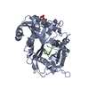
- Structure visualization
| Structure viewer | Molecule: MolmilJmol/JSmol |
|---|
- Downloads & links
-Download
| PDBx/mmCIF format | 5uli.cif.gz | 94.7 KB | Display | PDBx/mmCIF format |
|---|---|---|---|---|
| PDB format | pdb5uli.ent.gz | 70 KB | Display | PDB format |
| PDBx/mmJSON format | 5uli.json.gz | Tree view | PDBx/mmJSON format | |
| Others |  Other downloads Other downloads |
-Validation report
| Arichive directory | https://data.pdbj.org/pub/pdb/validation_reports/ul/5uliftp://data.pdbj.org/pub/pdb/validation_reports/ul/5uli | HTTPS FTP |
|---|
-Related structure data


-Links
 PDBj
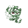
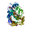
PDBj- Assembly
| Deposited unit | 
| ||||||||
|---|---|---|---|---|---|---|---|---|---|
| 1 |
| ||||||||
| Unit cell |
|
-Components
| #1: Protein | Mass: 43132.531 Da / Num. of mol.: 1 Source method: isolated from a genetically manipulated source Source: (gene. exp.) Mus musculus (house mouse) / Gene: Dxo, Dom3z, Ng6 / Plasmid: pet28a / Production host:  Escherichia coli (E. coli) / Strain (production host): BL21(DE3)Rosetta Escherichia coli (E. coli) / Strain (production host): BL21(DE3)RosettaReferences: UniProt: O70348, Hydrolases; Acting on ester bonds; Exoribonucleases producing 5'-phosphomonoesters, Hydrolases; Acting on acid anhydrides; In phosphorus-containing anhydrides |
|---|---|
| #2: Chemical | ChemComp-CA /   Mass: 40.078 Da / Num. of mol.: 1 / Source method: obtained synthetically / Formula: Ca Mass: 40.078 Da / Num. of mol.: 1 / Source method: obtained synthetically / Formula: Ca |
| #3: Chemical | ChemComp-0WD / [[(  Mass: 745.421 Da / Num. of mol.: 1 / Source method: obtained synthetically / Formula: C21H30N7O17P3 Mass: 745.421 Da / Num. of mol.: 1 / Source method: obtained synthetically / Formula: C21H30N7O17P3 |
| #4: Water | ChemComp-HOH / Water Mass: 18.015 Da / Num. of mol.: 151 / Source method: isolated from a natural source / Formula: H2O Mass: 18.015 Da / Num. of mol.: 151 / Source method: isolated from a natural source / Formula: H2O |
-Experimental details
-Experiment
| Experiment | Method: X-RAY DIFFRACTION / Number of used crystals: 1 |
|---|
- Sample preparation
| Crystal | Density Matthews: 2.61 Å3/Da / Density % sol: 52.89 % |
|---|---|
| Crystal grow | Temperature: 293 K / Method: vapor diffusion, hanging drop / pH: 7.5 / Details: 20% (w/v) PEG 3350 |
-Data collection
| Diffraction | Mean temperature: 100 K | ||||||||||||||||||||||||||||||||||||||||||||||||||||||||||||||||||||||||||||||||||||||||||||||||||||
|---|---|---|---|---|---|---|---|---|---|---|---|---|---|---|---|---|---|---|---|---|---|---|---|---|---|---|---|---|---|---|---|---|---|---|---|---|---|---|---|---|---|---|---|---|---|---|---|---|---|---|---|---|---|---|---|---|---|---|---|---|---|---|---|---|---|---|---|---|---|---|---|---|---|---|---|---|---|---|---|---|---|---|---|---|---|---|---|---|---|---|---|---|---|---|---|---|---|---|---|---|---|
| Diffraction source | Source: SYNCHROTRON / Site: APS  / Beamline: 24-ID-C / Wavelength: 0.979 Å / Beamline: 24-ID-C / Wavelength: 0.979 Å | ||||||||||||||||||||||||||||||||||||||||||||||||||||||||||||||||||||||||||||||||||||||||||||||||||||
| Detector | Type: DECTRIS PILATUS 6M / Detector: PIXEL / Date: Apr 11, 2016 | ||||||||||||||||||||||||||||||||||||||||||||||||||||||||||||||||||||||||||||||||||||||||||||||||||||
| Radiation | Protocol: SINGLE WAVELENGTH / Monochromatic (M) / Laue (L): M / Scattering type: x-ray | ||||||||||||||||||||||||||||||||||||||||||||||||||||||||||||||||||||||||||||||||||||||||||||||||||||
| Radiation wavelength | Wavelength: 0.979 Å / Relative weight: 1 | ||||||||||||||||||||||||||||||||||||||||||||||||||||||||||||||||||||||||||||||||||||||||||||||||||||
| Reflection | Resolution: 2.1→48.926 Å / Num. obs: 47716 / % possible obs: 98.2 % / Observed criterion σ(I): -3 / Redundancy: 2.902 % / Biso Wilson estimate: 26.96 Å2 / CC1/2: 0.997 / Rmerge(I) obs: 0.072 / Rrim(I) all: 0.088 / Χ2: 0.943 / Net I/σ(I): 12.06 / Num. measured all: 137567 | ||||||||||||||||||||||||||||||||||||||||||||||||||||||||||||||||||||||||||||||||||||||||||||||||||||
| Reflection shell | Diffraction-ID: 1
|
-Phasing
| Phasing | Method: molecular replacement | |||||||||
|---|---|---|---|---|---|---|---|---|---|---|
| Phasing MR |
|
- Processing
| Software |
| |||||||||||||||||||||||||||||||||||||||||||||||||||||||||||||||||||||||||||||
|---|---|---|---|---|---|---|---|---|---|---|---|---|---|---|---|---|---|---|---|---|---|---|---|---|---|---|---|---|---|---|---|---|---|---|---|---|---|---|---|---|---|---|---|---|---|---|---|---|---|---|---|---|---|---|---|---|---|---|---|---|---|---|---|---|---|---|---|---|---|---|---|---|---|---|---|---|---|---|
| Refinement | Method to determine structure: MOLECULAR REPLACEMENT / Resolution: 2.1→48.926 Å / SU ML: 0.21 / Cross valid method: FREE R-VALUE / σ(F): 1.14 / Phase error: 21.62 / Stereochemistry target values: ML
| |||||||||||||||||||||||||||||||||||||||||||||||||||||||||||||||||||||||||||||
| Solvent computation | Shrinkage radii: 0.9 Å / VDW probe radii: 1.11 Å / Solvent model: FLAT BULK SOLVENT MODEL | |||||||||||||||||||||||||||||||||||||||||||||||||||||||||||||||||||||||||||||
| Displacement parameters | Biso max: 91.64 Å2 / Biso mean: 30.9009 Å2 / Biso min: 13.7 Å2 | |||||||||||||||||||||||||||||||||||||||||||||||||||||||||||||||||||||||||||||
| Refinement step | Cycle: final / Resolution: 2.1→48.926 Å
| |||||||||||||||||||||||||||||||||||||||||||||||||||||||||||||||||||||||||||||
| Refine LS restraints |
| |||||||||||||||||||||||||||||||||||||||||||||||||||||||||||||||||||||||||||||
| LS refinement shell | Refine-ID: X-RAY DIFFRACTION / Rfactor Rfree error: 0 / Total num. of bins used: 10
|
