 Movie
Movie Controller

[English] 日本語
 Yorodumi
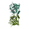
- PDB-5uc1: Structural Analysis of Glucocorticoid Receptor beta Ligand Bindin... -
+ Open data

- Basic information
| Entry | Database: PDB / ID: 5uc1 | ||||||
|---|---|---|---|---|---|---|---|
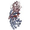
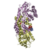
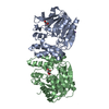
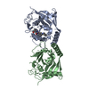
| Title | Structural Analysis of Glucocorticoid Receptor beta Ligand Binding Domain Complexed with Glucocorticoid Antagonist RU-486: Implication of Helix 12 in Antagonism | ||||||
 Components Components | Glucocorticoid receptor | ||||||
 Keywords Keywords | HORMONE RECEPTOR / Nuclear receptor | ||||||
| Function / homology |  Function and homology information Function and homology informationnuclear glucocorticoid receptor activity / steroid hormone binding / core promoter sequence-specific DNA binding / cellular response to transforming growth factor beta stimulus / cellular response to dexamethasone stimulus / Hsp90 protein binding / DNA-binding transcription repressor activity, RNA polymerase II-specific / positive regulation of miRNA transcription / spindle / nuclear receptor activity ...nuclear glucocorticoid receptor activity / steroid hormone binding / core promoter sequence-specific DNA binding / cellular response to transforming growth factor beta stimulus / cellular response to dexamethasone stimulus / Hsp90 protein binding / DNA-binding transcription repressor activity, RNA polymerase II-specific / positive regulation of miRNA transcription / spindle / nuclear receptor activity / chromatin organization / DNA-binding transcription activator activity, RNA polymerase II-specific / nuclear speck / RNA polymerase II cis-regulatory region sequence-specific DNA binding / protein kinase binding / protein-containing complex / mitochondrion / zinc ion binding / cytosolSimilarity search - Function | ||||||
| Biological species |  Heterocephalus glaber (naked mole-rat) Heterocephalus glaber (naked mole-rat) | ||||||
| Method | X-RAY DIFFRACTION / SYNCHROTRON / MOLECULAR REPLACEMENT / molecular replacement / Resolution: 2.351 Å | ||||||
 Authors Authors | Pedersen, L.C. / Min, J. | ||||||
 Citation Citation | Journal: Mol. Cell. Biol. / Year: 2018 Title: Probing Dominant Negative Behavior of Glucocorticoid Receptor beta through a Hybrid Structural and Biochemical Approach. Authors: Min, J. / Perera, L. / Krahn, J.M. / Jewell, C.M. / Moon, A.F. / Cidlowski, J.A. / Pedersen, L.C. | ||||||
| History |
|
- Structure visualization
| Structure viewer | Molecule: MolmilJmol/JSmol |
|---|
- Downloads & links
-Download
| PDBx/mmCIF format | 5uc1.cif.gz | 102 KB | Display | PDBx/mmCIF format |
|---|---|---|---|---|
| PDB format | pdb5uc1.ent.gz | 75.9 KB | Display | PDB format |
| PDBx/mmJSON format | 5uc1.json.gz | Tree view | PDBx/mmJSON format | |
| Others |  Other downloads Other downloads |
-Validation report
| Arichive directory | https://data.pdbj.org/pub/pdb/validation_reports/uc/5uc1ftp://data.pdbj.org/pub/pdb/validation_reports/uc/5uc1 | HTTPS FTP |
|---|
-Related structure data
| Related structure data |  1m2zS S: Starting model for refinement |
|---|---|
| Similar structure data |


-Links
 PDBj
PDBj
- Assembly
| Deposited unit | 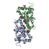
| ||||||||
|---|---|---|---|---|---|---|---|---|---|
| 1 |
| ||||||||
| Unit cell |
| ||||||||
| Details | THE AUTHOR STATES THAT THE BIOLOGICAL UNIT OF THIS PROTEIN IS UNKNOWN. |
-Components
-Protein , 1 types, 2 molecules AB
| #1: Protein | Mass: 25634.705 Da / Num. of mol.: 2 Source method: isolated from a genetically manipulated source Source: (gene. exp.) Heterocephalus glaber (naked mole-rat) / Gene: GW7_10599 / Production host:  Escherichia coli (E. coli) / References: UniProt: G5AQS2*PLUS Escherichia coli (E. coli) / References: UniProt: G5AQS2*PLUS |
|---|
-Non-polymers , 6 types, 36 molecules 
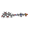


| #2: Chemical | Mifepristone Mass: 429.594 Da / Num. of mol.: 2 / Source method: obtained synthetically / Formula: C29H35NO2 / Comment: medication*YM Mass: 429.594 Da / Num. of mol.: 2 / Source method: obtained synthetically / Formula: C29H35NO2 / Comment: medication*YM#3: Chemical | ChemComp-CPS / CHAPS detergent Mass: 614.877 Da / Num. of mol.: 4 / Source method: obtained synthetically / Formula: C32H58N2O7S / Comment: detergent*YM Mass: 614.877 Da / Num. of mol.: 4 / Source method: obtained synthetically / Formula: C32H58N2O7S / Comment: detergent*YM#4: Chemical | ChemComp-CL / Chloride Mass: 35.453 Da / Num. of mol.: 4 / Source method: obtained synthetically / Formula: Cl Mass: 35.453 Da / Num. of mol.: 4 / Source method: obtained synthetically / Formula: Cl#5: Chemical | 2-Methyl-2,4-pentanediol Mass: 118.174 Da / Num. of mol.: 2 / Source method: obtained synthetically / Formula: C6H14O2 / Comment: precipitant*YM Mass: 118.174 Da / Num. of mol.: 2 / Source method: obtained synthetically / Formula: C6H14O2 / Comment: precipitant*YM#6: Chemical | ChemComp-EDO / Ethylene glycol Mass: 62.068 Da / Num. of mol.: 5 / Source method: obtained synthetically / Formula: C2H6O2 Mass: 62.068 Da / Num. of mol.: 5 / Source method: obtained synthetically / Formula: C2H6O2#7: Water | ChemComp-HOH / | WaterMass: 18.015 Da / Num. of mol.: 19 / Source method: isolated from a natural source / Formula: H2O |
|---|
-Experimental details
-Experiment
| Experiment | Method: X-RAY DIFFRACTION / Number of used crystals: 1 |
|---|
- Sample preparation
| Crystal | Density Matthews: 2.74 Å3/Da / Density % sol: 55.06 % |
|---|---|
| Crystal grow | Temperature: 277 K / Method: vapor diffusion, sitting drop / Details: 0.1 M MES pH 5.0 and 20 % MPD (v/v) |
-Data collection
| Diffraction | Mean temperature: 100 K |
|---|---|
| Diffraction source | Source: SYNCHROTRON / Site: APS  / Beamline: 22-ID / Wavelength: 1 Å / Beamline: 22-ID / Wavelength: 1 Å |
| Detector | Type: MARMOSAIC 300 mm CCD / Detector: CCD / Date: Mar 25, 2016 |
| Radiation | Protocol: SINGLE WAVELENGTH / Monochromatic (M) / Laue (L): M / Scattering type: x-ray |
| Radiation wavelength | Wavelength: 1 Å / Relative weight: 1 |
| Reflection | Resolution: 2.35→50 Å / Num. obs: 20767 / % possible obs: 98 % / Redundancy: 5.8 % / Rmerge(I) obs: 0.069 / Net I/σ(I): 28.6 |
| Reflection shell | Rmerge(I) obs: 0.453 |
-Phasing
| Phasing | Method: molecular replacement |
|---|
- Processing
| Software |
| ||||||||||||||||||||||||||||||||||||||||||||||||||||||||
|---|---|---|---|---|---|---|---|---|---|---|---|---|---|---|---|---|---|---|---|---|---|---|---|---|---|---|---|---|---|---|---|---|---|---|---|---|---|---|---|---|---|---|---|---|---|---|---|---|---|---|---|---|---|---|---|---|---|
| Refinement | Method to determine structure: MOLECULAR REPLACEMENT Starting model: 1M2Z Resolution: 2.351→35.094 Å / SU ML: 0.32 / Cross valid method: FREE R-VALUE / σ(F): 1.35 / Phase error: 31.88
| ||||||||||||||||||||||||||||||||||||||||||||||||||||||||
| Solvent computation | Shrinkage radii: 0.9 Å / VDW probe radii: 1.11 Å | ||||||||||||||||||||||||||||||||||||||||||||||||||||||||
| Displacement parameters | Biso max: 105.6 Å2 / Biso mean: 57.6788 Å2 / Biso min: 34.52 Å2 | ||||||||||||||||||||||||||||||||||||||||||||||||||||||||
| Refinement step | Cycle: final / Resolution: 2.351→35.094 Å
| ||||||||||||||||||||||||||||||||||||||||||||||||||||||||
| Refine LS restraints |
| ||||||||||||||||||||||||||||||||||||||||||||||||||||||||
| LS refinement shell | Refine-ID: X-RAY DIFFRACTION / Rfactor Rfree error: 0 / Total num. of bins used: 7
|
