 Movie
Movie Controller


+ Open data

- Basic information
| Entry | Database: PDB / ID: 5u4z | ||||||
|---|---|---|---|---|---|---|---|
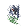
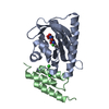
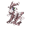
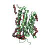
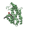
| Title | Crystal structure of citrus MAF1 in space group P 31 2 1 | ||||||
 Components Components | Repressor of RNA polymerase III transcription | ||||||
 Keywords Keywords |  TRANSCRIPTION / repressor of RNA polymerase III TRANSCRIPTION / repressor of RNA polymerase III | ||||||
| Function / homology | Repressor of RNA polymerase III transcription Maf1 / Repressor of RNA polymerase III transcription Maf1 superfamily / Maf1 regulator / negative regulation of transcription by RNA polymerase III / nucleus / Repressor of RNA polymerase III transcription Function and homology information Function and homology information | ||||||
| Biological species |  Citrus sinensis (sweet orange) Citrus sinensis (sweet orange) | ||||||
| Method | X-RAY DIFFRACTION / SYNCHROTRON / SAD / Resolution: 2.85 Å | ||||||
 Authors Authors | Soprano, A.S. / Giuseppe, P.O. / Nascimento, A.F.Z. / Benedetti, C.E. / Murakami, M.T. | ||||||
| Funding support |  Brazil, 1items Brazil, 1items
| ||||||
 Citation Citation | Journal: To Be Published Title: Crystal structure of citrus MAF1 in space group P 31 2 1 Authors: Soprano, A.S. / Giuseppe, P.O. / Nascimento, A.F.Z. / Benedetti, C.E. / Murakami, M.T. | ||||||
| History |
|
- Structure visualization
| Structure viewer | Molecule: MolmilJmol/JSmol |
|---|
- Downloads & links
-Download
| PDBx/mmCIF format | 5u4z.cif.gz | 86.5 KB | Display | PDBx/mmCIF format |
|---|---|---|---|---|
| PDB format | pdb5u4z.ent.gz | 67.5 KB | Display | PDB format |
| PDBx/mmJSON format | 5u4z.json.gz | Tree view | PDBx/mmJSON format | |
| Others |  Other downloads Other downloads |
-Validation report
| Arichive directory | https://data.pdbj.org/pub/pdb/validation_reports/u4/5u4zftp://data.pdbj.org/pub/pdb/validation_reports/u4/5u4z | HTTPS FTP |
|---|
-Related structure data
| Similar structure data |
|---|


-Links
 PDBj
PDBj- Assembly
| Deposited unit | 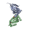
| ||||||||
|---|---|---|---|---|---|---|---|---|---|
| 1 | 
| ||||||||
| 2 | 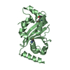
| ||||||||
| Unit cell |
|
-Components
| #1: Protein | Mass: 31098.385 Da / Num. of mol.: 2 Source method: isolated from a genetically manipulated source Source: (gene. exp.) Citrus sinensis (sweet orange) / Gene: MAF1 / Plasmid: pET28a / Production host:  Escherichia coli (E. coli) / References: UniProt: G9I821 Escherichia coli (E. coli) / References: UniProt: G9I821#2: Chemical | Sulfate  Mass: 96.063 Da / Num. of mol.: 2 / Source method: obtained synthetically / Formula: SO4 Mass: 96.063 Da / Num. of mol.: 2 / Source method: obtained synthetically / Formula: SO4#3: Water | ChemComp-HOH / | Water Mass: 18.015 Da / Num. of mol.: 1 / Source method: isolated from a natural source / Formula: H2O Mass: 18.015 Da / Num. of mol.: 1 / Source method: isolated from a natural source / Formula: H2O |
|---|
-Experimental details
-Experiment
| Experiment | Method: X-RAY DIFFRACTION / Number of used crystals: 1 |
|---|
- Sample preparation
| Crystal | Density Matthews: 3.13 Å3/Da / Density % sol: 60.76 % |
|---|---|
| Crystal grow | Temperature: 291.15 K / Method: vapor diffusion / pH: 5.6 Details: 1 M ammonium di-hydrogen phosphate, 0.1 M Tris-sodium pH 5.6, 10% glycerol |
-Data collection
| Diffraction | Mean temperature: 100 K | ||||||||||||||||||||||||||||||||||||||||||||||||||
|---|---|---|---|---|---|---|---|---|---|---|---|---|---|---|---|---|---|---|---|---|---|---|---|---|---|---|---|---|---|---|---|---|---|---|---|---|---|---|---|---|---|---|---|---|---|---|---|---|---|---|---|
| Diffraction source | Source: SYNCHROTRON / Site: ALBA  / Beamline: XALOC / Wavelength: 0.979 Å / Beamline: XALOC / Wavelength: 0.979 Å | ||||||||||||||||||||||||||||||||||||||||||||||||||
| Detector | Type: DECTRIS PILATUS 6M / Detector: PIXEL / Date: Nov 16, 2015 | ||||||||||||||||||||||||||||||||||||||||||||||||||
| Radiation | Protocol: SINGLE WAVELENGTH / Monochromatic (M) / Laue (L): M / Scattering type: x-ray | ||||||||||||||||||||||||||||||||||||||||||||||||||
| Radiation wavelength | Wavelength: 0.979 Å / Relative weight: 1 | ||||||||||||||||||||||||||||||||||||||||||||||||||
| Reflection | Resolution: 2.85→47.086 Å / Num. obs: 35279 / % possible obs: 100 % / Observed criterion σ(I): -3 / Redundancy: 9.4 % / Biso Wilson estimate: 69.534 Å2 / CC1/2: 0.997 / Rmerge(I) obs: 0.214 / Rrim(I) all: 0.225 / Χ2: 1.468 / Net I/σ(I): 10.94 / Num. measured all: 372748 | ||||||||||||||||||||||||||||||||||||||||||||||||||
| Reflection shell |
|
- Processing
| Software |
| |||||||||||||||||||||||||||||||||||||||||||||||||||||||||||||||||||||||||||
|---|---|---|---|---|---|---|---|---|---|---|---|---|---|---|---|---|---|---|---|---|---|---|---|---|---|---|---|---|---|---|---|---|---|---|---|---|---|---|---|---|---|---|---|---|---|---|---|---|---|---|---|---|---|---|---|---|---|---|---|---|---|---|---|---|---|---|---|---|---|---|---|---|---|---|---|---|
| Refinement | Method to determine structure: SAD / Resolution: 2.85→20 Å / Cor.coef. Fo:Fc: 0.956 / Cor.coef. Fo:Fc free: 0.935 / WRfactor Rfree: 0.2009 / WRfactor Rwork: 0.1655 / FOM work R set: 0.7799 / SU B: 14.493 / SU ML: 0.258 / SU R Cruickshank DPI: 0.4087 / SU Rfree: 0.2769 / Cross valid method: THROUGHOUT / σ(F): 0 / ESU R: 0.409 / ESU R Free: 0.277 / Stereochemistry target values: MAXIMUM LIKELIHOOD Details: HYDROGENS HAVE BEEN ADDED IN THE RIDING POSITIONS U VALUES : REFINED INDIVIDUALLY
| |||||||||||||||||||||||||||||||||||||||||||||||||||||||||||||||||||||||||||
| Solvent computation | Ion probe radii: 0.8 Å / Shrinkage radii: 0.8 Å / VDW probe radii: 1.2 Å / Solvent model: MASK | |||||||||||||||||||||||||||||||||||||||||||||||||||||||||||||||||||||||||||
| Displacement parameters | Biso max: 153.12 Å2 / Biso mean: 81.165 Å2 / Biso min: 50.75 Å2
| |||||||||||||||||||||||||||||||||||||||||||||||||||||||||||||||||||||||||||
| Refinement step | Cycle: final / Resolution: 2.85→20 Å
| |||||||||||||||||||||||||||||||||||||||||||||||||||||||||||||||||||||||||||
| Refine LS restraints |
| |||||||||||||||||||||||||||||||||||||||||||||||||||||||||||||||||||||||||||
| LS refinement shell | Resolution: 2.85→2.922 Å / Total num. of bins used: 20
|
