 Movie
Movie Controller

[English] 日本語
 Yorodumi
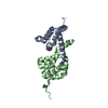
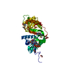
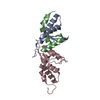
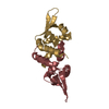
- PDB-5hyb: Crystal structure of myristoylated Y81A mutant MMTV matrix protein -
+ Open data

- Basic information
| Entry | Database: PDB / ID: 5hyb | |||||||||||||||||||||
|---|---|---|---|---|---|---|---|---|---|---|---|---|---|---|---|---|---|---|---|---|---|---|
| Title | Crystal structure of myristoylated Y81A mutant MMTV matrix protein | |||||||||||||||||||||
 Components Components | Matrix Protein Viral matrix protein Viral matrix protein | |||||||||||||||||||||
 Keywords Keywords | VIRAL PROTEIN / Myristoylated protein / MMTV mutant | |||||||||||||||||||||
| Function / homology |  Function and homology information Function and homology informationviral budding via host ESCRT complex / viral nucleocapsid / structural constituent of virion / nucleotide binding / DNA binding / zinc ion bindingSimilarity search - Function | |||||||||||||||||||||
| Biological species |  Mouse mammary tumor virus Mouse mammary tumor virus | |||||||||||||||||||||
| Method | X-RAY DIFFRACTION / SYNCHROTRON / FOURIER SYNTHESIS / Resolution: 1.94 Å | |||||||||||||||||||||
 Authors Authors | Brynda, J. / Dostal, J. / Zabransky, A. / Dolezal, M. | |||||||||||||||||||||
| Funding support |  Czech Republic, 6items Czech Republic, 6items
| |||||||||||||||||||||
 Citation Citation | Journal: To Be Published Title: Crystal structure of myristoylated Y81A mutant MMTV matrix protein Authors: Brynda, J. / Dostal, J. / Zabransky, A. / Dolezal, M. | |||||||||||||||||||||
| History |
|
- Structure visualization
| Structure viewer | Molecule: MolmilJmol/JSmol |
|---|
- Downloads & links
-Download
| PDBx/mmCIF format | 5hyb.cif.gz | 49.1 KB | Display | PDBx/mmCIF format |
|---|---|---|---|---|
| PDB format | pdb5hyb.ent.gz | 38.5 KB | Display | PDB format |
| PDBx/mmJSON format | 5hyb.json.gz | Tree view | PDBx/mmJSON format | |
| Others |  Other downloads Other downloads |
-Validation report
| Arichive directory | https://data.pdbj.org/pub/pdb/validation_reports/hy/5hybftp://data.pdbj.org/pub/pdb/validation_reports/hy/5hyb | HTTPS FTP |
|---|
-Related structure data
| Related structure data | |
|---|---|
| Similar structure data |


-Links
 PDBj
PDBj


- Assembly
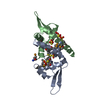
| Deposited unit | 
| ||||||||
|---|---|---|---|---|---|---|---|---|---|
| 1 | 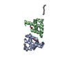
| ||||||||
| 2 | 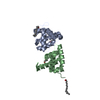
| ||||||||
| Unit cell |
|
-Components
| #1: Protein | Viral matrix protein / Gag polyprotein Mass: 10396.958 Da / Num. of mol.: 2 / Mutation: Y81A Source method: isolated from a genetically manipulated source Source: (gene. exp.) Mouse mammary tumor virus (strain BR6) / Gene: gag / Production host:  Escherichia coli BL21(DE3) (bacteria) / References: UniProt: P10258 Escherichia coli BL21(DE3) (bacteria) / References: UniProt: P10258#2: Chemical | Myristic acid  Mass: 228.371 Da / Num. of mol.: 2 / Source method: obtained synthetically / Formula: C14H28O2 Mass: 228.371 Da / Num. of mol.: 2 / Source method: obtained synthetically / Formula: C14H28O2#3: Chemical | Diethylene glycol  Mass: 106.120 Da / Num. of mol.: 2 / Source method: obtained synthetically / Formula: C4H10O3 Mass: 106.120 Da / Num. of mol.: 2 / Source method: obtained synthetically / Formula: C4H10O3#4: Water | ChemComp-HOH / | Water Mass: 18.015 Da / Num. of mol.: 89 / Source method: isolated from a natural source / Formula: H2O Mass: 18.015 Da / Num. of mol.: 89 / Source method: isolated from a natural source / Formula: H2O |
|---|
-Experimental details
-Experiment
| Experiment | Method: X-RAY DIFFRACTION / Number of used crystals: 1 |
|---|
- Sample preparation
| Crystal | Density Matthews: 2.38 Å3/Da / Density % sol: 48.23 % |
|---|---|
| Crystal grow | Temperature: 292 K / Method: vapor diffusion, sitting drop / Details: 0.1M Tris-HCl pH 8.5, 25% (v/v) PEG 550 MME |
-Data collection
| Diffraction | Mean temperature: 100 K |
|---|---|
| Diffraction source | Source: SYNCHROTRON / Site: BESSY  / Beamline: 14.2 / Wavelength: 0.91841 Å / Beamline: 14.2 / Wavelength: 0.91841 Å |
| Detector | Type: MARMOSAIC 225 mm CCD / Detector: CCD / Date: May 29, 2013 |
| Radiation | Monochromator: Optics / Protocol: SINGLE WAVELENGTH / Monochromatic (M) / Laue (L): M / Scattering type: x-ray |
| Radiation wavelength | Wavelength: 0.91841 Å / Relative weight: 1 |
| Reflection | Resolution: 1.93→50 Å / Num. obs: 13822 / % possible obs: 89.7 % / Redundancy: 6 % / Biso Wilson estimate: 35.3 Å2 / CC1/2: 0.999 / Net I/σ(I): 15.6 |
| Reflection shell | Resolution: 1.93→2.05 Å / Redundancy: 6 % / Rmerge(I) obs: 0.906 / Mean I/σ(I) obs: 2.01 / % possible all: 80.1 |
- Processing
| Software |
| ||||||||||||||||||||||||||||||||||||||||||||||||||||||||||||
|---|---|---|---|---|---|---|---|---|---|---|---|---|---|---|---|---|---|---|---|---|---|---|---|---|---|---|---|---|---|---|---|---|---|---|---|---|---|---|---|---|---|---|---|---|---|---|---|---|---|---|---|---|---|---|---|---|---|---|---|---|---|
| Refinement | Method to determine structure: FOURIER SYNTHESIS / Resolution: 1.94→45.95 Å / Cor.coef. Fo:Fc: 0.945 / Cor.coef. Fo:Fc free: 0.924 / SU B: 5.417 / SU ML: 0.149 / SU R Cruickshank DPI: 0.2255 / Cross valid method: THROUGHOUT / σ(F): 0 / ESU R: 0.225 / ESU R Free: 0.202 Details: HYDROGENS HAVE BEEN ADDED IN THE RIDING POSITIONS U VALUES : REFINED INDIVIDUALLY
| ||||||||||||||||||||||||||||||||||||||||||||||||||||||||||||
| Solvent computation | Ion probe radii: 0.8 Å / Shrinkage radii: 0.8 Å / VDW probe radii: 1.2 Å | ||||||||||||||||||||||||||||||||||||||||||||||||||||||||||||
| Displacement parameters | Biso max: 89.9 Å2 / Biso mean: 27.27 Å2 / Biso min: 12.67 Å2
| ||||||||||||||||||||||||||||||||||||||||||||||||||||||||||||
| Refinement step | Cycle: final / Resolution: 1.94→45.95 Å
| ||||||||||||||||||||||||||||||||||||||||||||||||||||||||||||
| Refine LS restraints |
| ||||||||||||||||||||||||||||||||||||||||||||||||||||||||||||
| LS refinement shell | Resolution: 1.942→1.992 Å / Total num. of bins used: 20
|
